Copyright
©The Author(s) 2025.
World J Gastrointest Oncol. Jun 15, 2025; 17(6): 106848
Published online Jun 15, 2025. doi: 10.4251/wjgo.v17.i6.106848
Published online Jun 15, 2025. doi: 10.4251/wjgo.v17.i6.106848
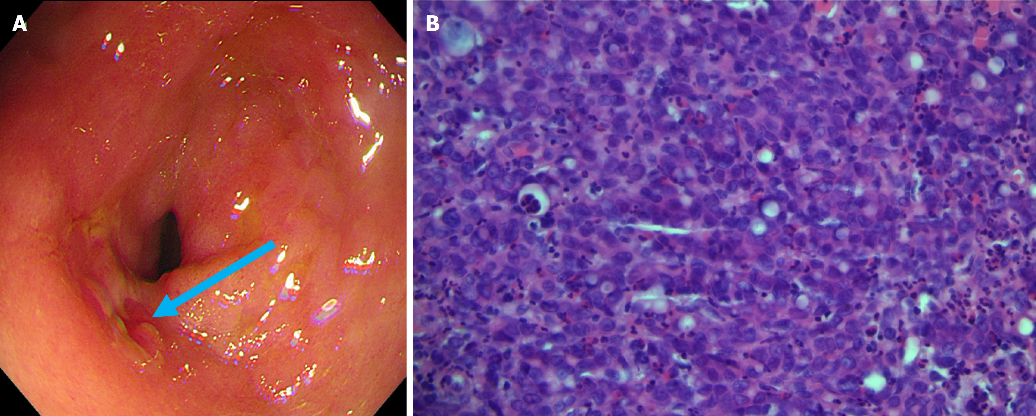
Figure 4 Patient’s gastroscopy and pathology images.
A: Gastric antral mucosa with scattered erythematous erosions, and a 1.0 cm erosion in the prepyloric area (blue arrow); B: Pathology showed the gastric antrum lesion to be consistent with poorly differentiated adenocarcinoma (hematoxylin and eosin 400 ×).
- Citation: Jin CB, Li YS, Zhang J, Wu J, Tao WJ. Extensive bone metastases from an occult gastric primary: A case report. World J Gastrointest Oncol 2025; 17(6): 106848
- URL: https://www.wjgnet.com/1948-5204/full/v17/i6/106848.htm
- DOI: https://dx.doi.org/10.4251/wjgo.v17.i6.106848


