Copyright
©The Author(s) 2025.
World J Gastrointest Oncol. May 15, 2025; 17(5): 104011
Published online May 15, 2025. doi: 10.4251/wjgo.v17.i5.104011
Published online May 15, 2025. doi: 10.4251/wjgo.v17.i5.104011
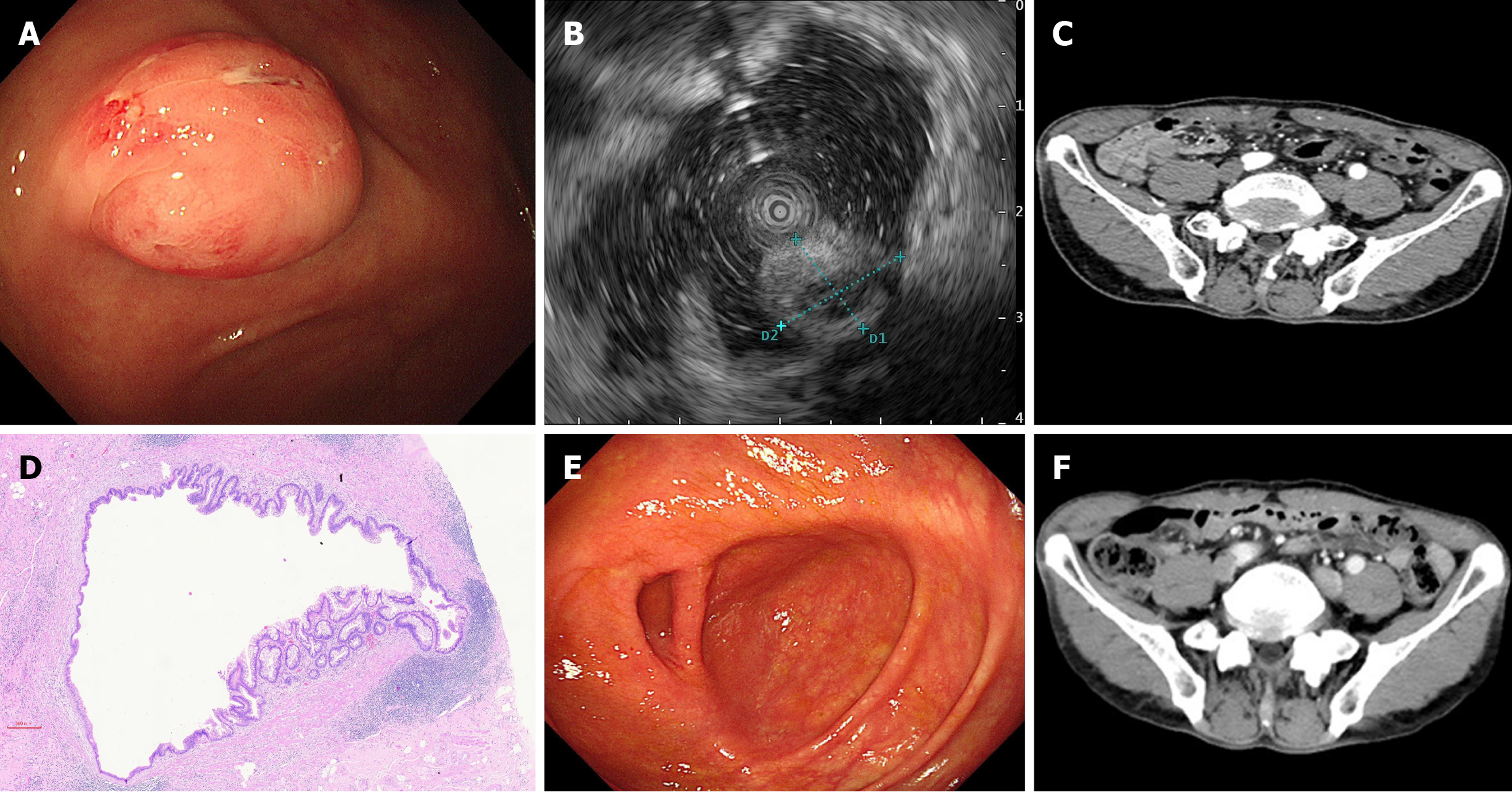
Figure 1 Clinical data of the patient with a low-grade appendiceal mucinous neoplasm at the appendiceal orifice.
A: Normal colonoscopy showed a submucosal protrusion at the appendiceal orifice; B: Ultrasound colonoscopy image; C: Abdominal computed tomography enhancement; D: Pathology suggested a low-grade mucinous cystadenoma of the appendix with no involvement of the margins; E: Repeat colonoscopy 18 months after surgery revealed no significant abnormality at the appendiceal orifice; F: Computed tomography enhancement of the abdomen suggested postoperative changes in the appendix.
Figure 2 Double purse-string suture technique.
A: A purse-string suture was created approximately 1.0 cm below the swelling; B: A cut was made 0.5 cm from the ligature line; C: A second purse-string suture was performed 1.0 cm from the ligature line; D: The remaining end was embedded into the purse-string suture.
- Citation: Liu D, Xing YL, Chen D. Low-grade appendiceal mucinous neoplasm at appendiceal orifice treated via appendectomy with double purse-string suture method: A case report. World J Gastrointest Oncol 2025; 17(5): 104011
- URL: https://www.wjgnet.com/1948-5204/full/v17/i5/104011.htm
- DOI: https://dx.doi.org/10.4251/wjgo.v17.i5.104011


