Copyright
©The Author(s) 2024.
World J Gastrointest Oncol. Feb 15, 2024; 16(2): 550-556
Published online Feb 15, 2024. doi: 10.4251/wjgo.v16.i2.550
Published online Feb 15, 2024. doi: 10.4251/wjgo.v16.i2.550
Figure 1 Abdominal enhanced computed tomography images.
A: Arterial phase; B: Portal vein phase; C: Coronal plane. The liver is significantly enlarged. The contour is irregular, the density of the parenchyma is reduced and uneven, and multiple nodular and punctate enhancements can be seen during the arterial phase. The spleen is enlarged.
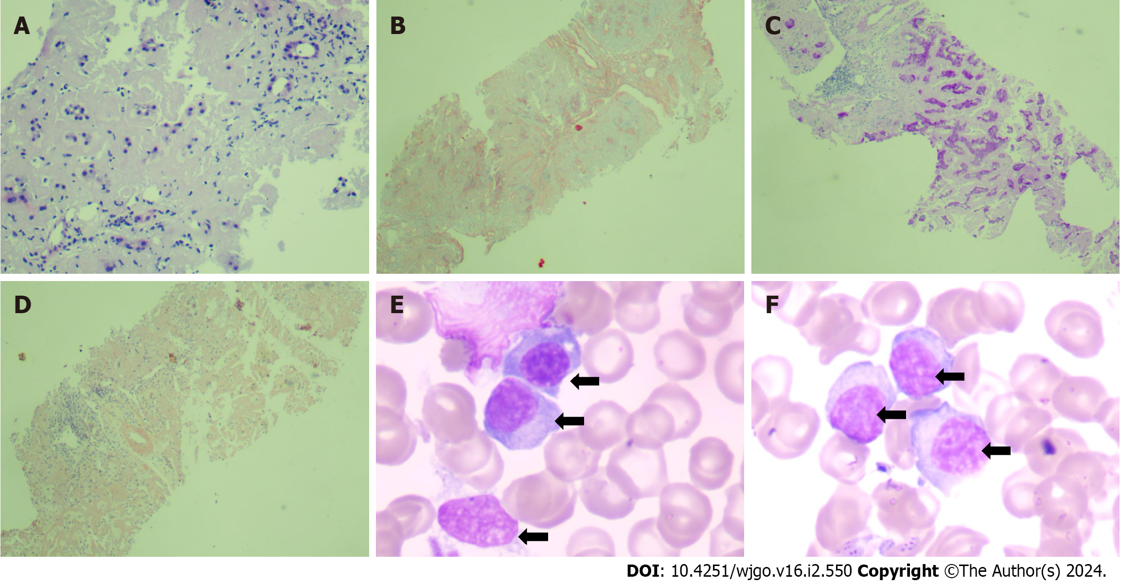
Figure 2 Liver pathology and bone marrow aspiration smear.
A: Hematoxylin-eosin staining (100 ×) showed deposition of pink fine-grained amorphous material in hepatic lobule and portal area, compression and deformation of liver plates, and atrophy of hepatocytes; B: Masson staining (40 ×) showed mild peri-sinusoidal fibrosis; C: Periodic acid-Schiff staining (40 ×) showed significant atrophy of liver cells with a few residual liver cells; D: Congo red staining (40 ×) indicated brick-red material deposition between liver cells; E and F: Bone marrow aspiration smear (100 ×) showed abnormal proliferation of plasma cell system. The cells are moderate-sized, the nucleus is large and eccentric, round or oval, the nuclear chromatin is fine, the cytoplasm is rich, blue, and foamy (black arrows).
- Citation: Zhang X, Tang F, Gao YY, Song DZ, Liang J. Hepatomegaly and jaundice as the presenting symptoms of systemic light-chain amyloidosis: A case report. World J Gastrointest Oncol 2024; 16(2): 550-556
- URL: https://www.wjgnet.com/1948-5204/full/v16/i2/550.htm
- DOI: https://dx.doi.org/10.4251/wjgo.v16.i2.550


