Copyright
©The Author(s) 2021.
World J Gastrointest Oncol. Jun 15, 2021; 13(6): 462-471
Published online Jun 15, 2021. doi: 10.4251/wjgo.v13.i6.462
Published online Jun 15, 2021. doi: 10.4251/wjgo.v13.i6.462
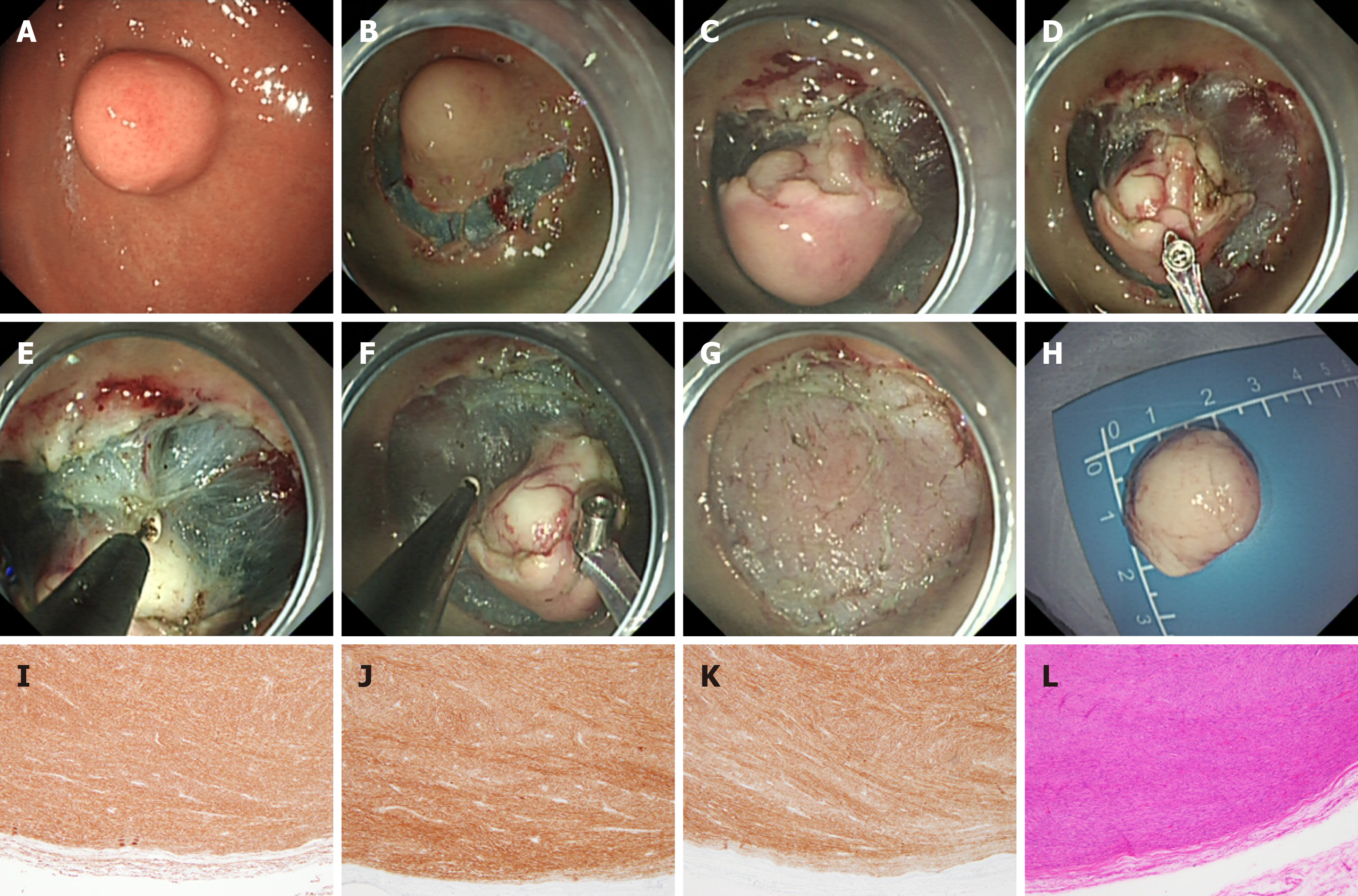
Figure 1 Endoscopic submucosal dissection treatment of gastric stromal tumor.
A: Gastric stromal tumor (GST) in the fundus of the stomach; B: Submucosal injection around the GST was performed with an injection needle, and then a submucosal incision was made; C: The tumor (white) can be seen after peripheral submucosal separation; D: Traction of the tumor by the clip-and-snare method to expose its root; E and F: An IT knife was used to separate the root of the tumor; G: Wound surface after tumor resection; H: Complete resection of the tumor; I-K: CD34 (I), CD117 (J), and DOG-1 (K) were all expressed; L: Mitotic figure count ≤ 5 per 50 high-power fields.
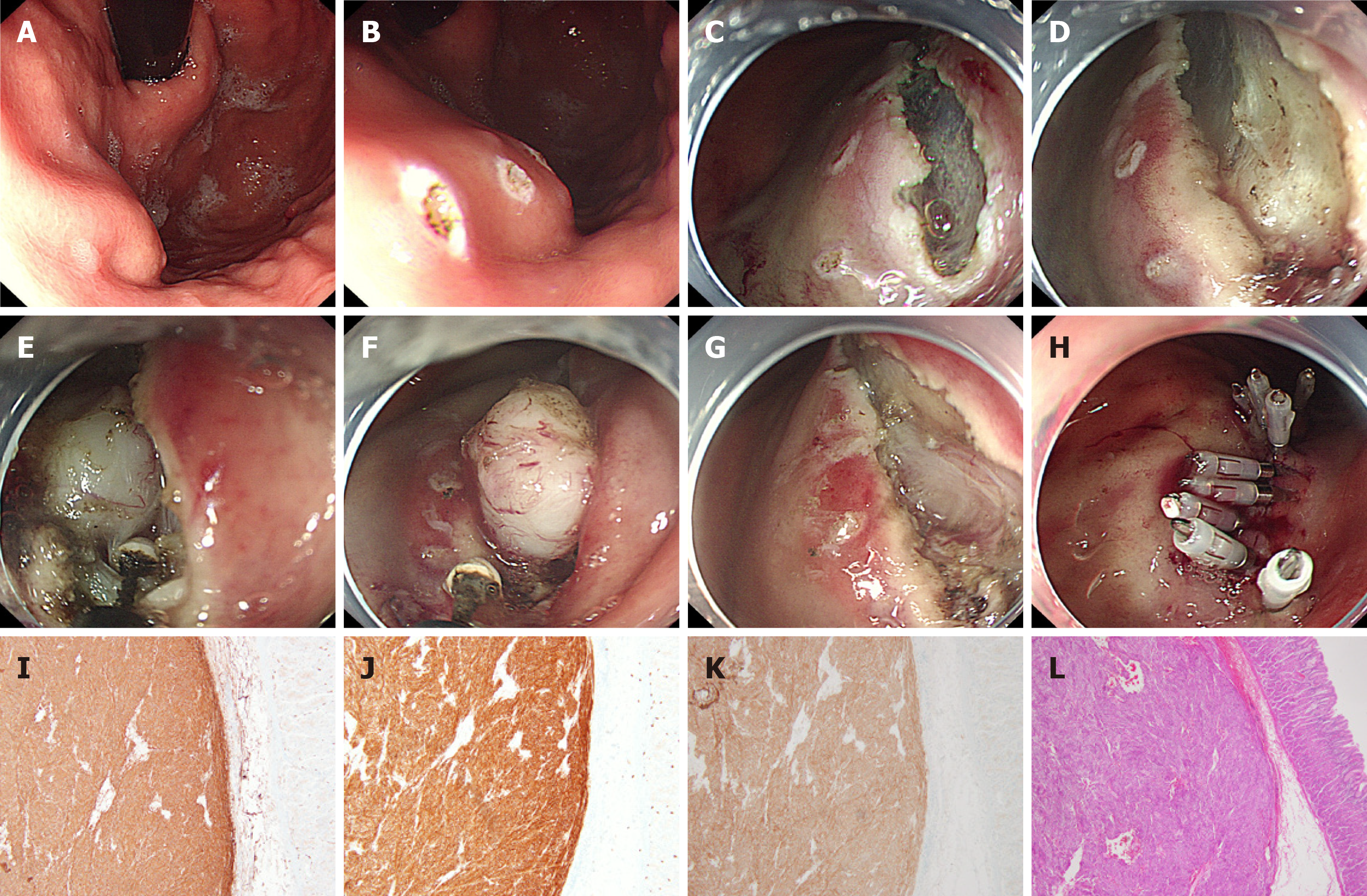
Figure 2 Endoscopic submucosal excavation treatment of gastric stromal tumor.
A: Gastric stromal tumor (GST) at the junction of the gastric body and fundus; B: “Linear” electrocoagulation labeling of the GST mucosa; C and D: The tumor (white) was observed after linear incision of the GST surface mucosa and submucosa; E and F: An IT knife was used to separate the root of the tumor; G: Wound surface after tumor resection; H: Titanium clip sealing of the wound; I-K: CD34 (I), CD117 (J), and DOG-1 (K) were all expressed; L: Mitotic figure count ≤ 5/50 high-power fields.
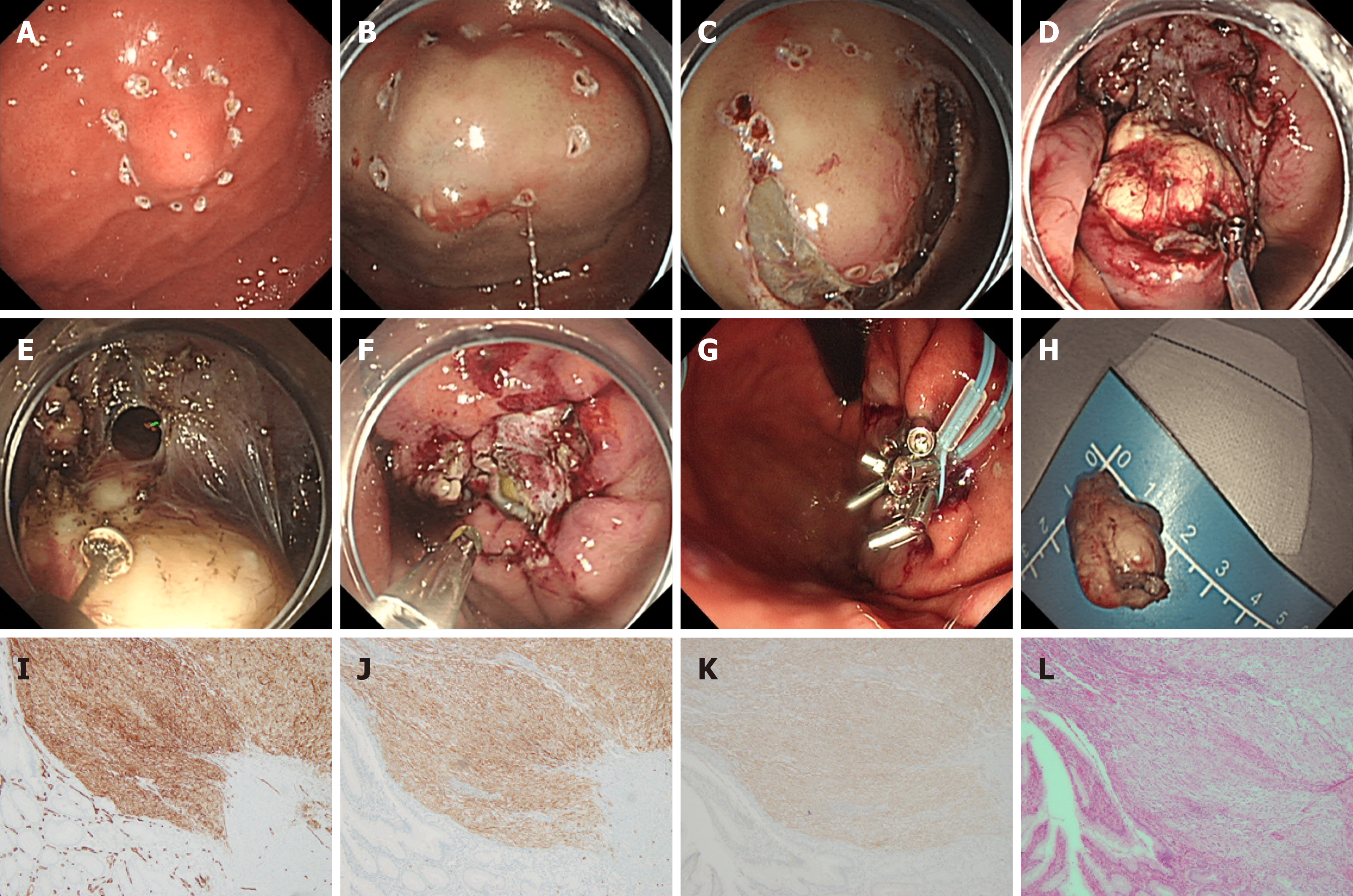
Figure 3 Endoscopic full-thickness resection treatment of gastric stromal tumor.
A: Gastric stromal tumor (GST) of the gastric body near the fundus; B: Submucosal injection around the GST was performed by injection needle; C: Circumferential incision of the submucosa around the tumor; D: Traction of the exposed tumor with the clip-and-snare method; E: An IT knife was used to separate the root of the tumor, and the local full-thickness gastric wall was cut open; F: After full-layer resection, the wound was treated with hot biopsy forceps for hot coagulation and hemostasis; G: After tumor resection, the wound was sealed with a titanium clip and a nylon ring for a purse pocket suture; H: Complete resection of the tumor body for examination; I-K: CD34 (I), CD117 (J), and DOG-1 (K) were all expressed; L: Mitotic figure count ≤ 5/50 high-power fields.
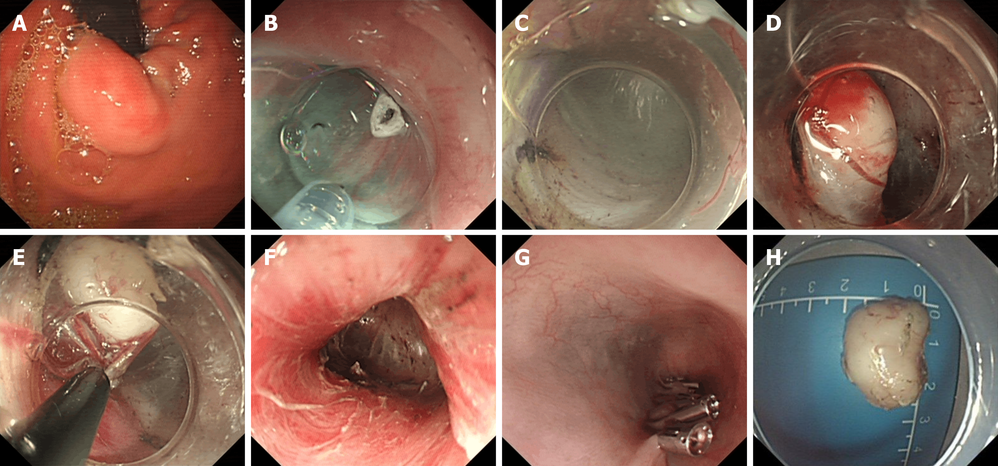
Figure 4 Submucosal-tunneling endoscopic resection treatment of gastric stromal tumor.
A: Gastric stromal tumor (GST) at the esophagogastric junction; B: Submucosal injection was initiated by needle in the esophagus about 5 cm away from the GST; C and D: The esophageal mucosa was cut open to establish a submucosal tunnel to the tumor; E: An IT knife was used to separate the root of the tumor; F: Heat coagulation and hemostatic therapy on wound surface after tumor resection; G: The opening of the esophageal tunnel was sealed with titanium clamps; H: Complete resection of the tumor.
- Citation: Chen ZM, Peng MS, Wang LS, Xu ZL. Efficacy and safety of endoscopic resection in treatment of small gastric stromal tumors: A state-of-the-art review. World J Gastrointest Oncol 2021; 13(6): 462-471
- URL: https://www.wjgnet.com/1948-5204/full/v13/i6/462.htm
- DOI: https://dx.doi.org/10.4251/wjgo.v13.i6.462


