Copyright
©The Author(s) 2021.
World J Gastrointest Oncol. Dec 15, 2021; 13(12): 2180-2189
Published online Dec 15, 2021. doi: 10.4251/wjgo.v13.i12.2180
Published online Dec 15, 2021. doi: 10.4251/wjgo.v13.i12.2180
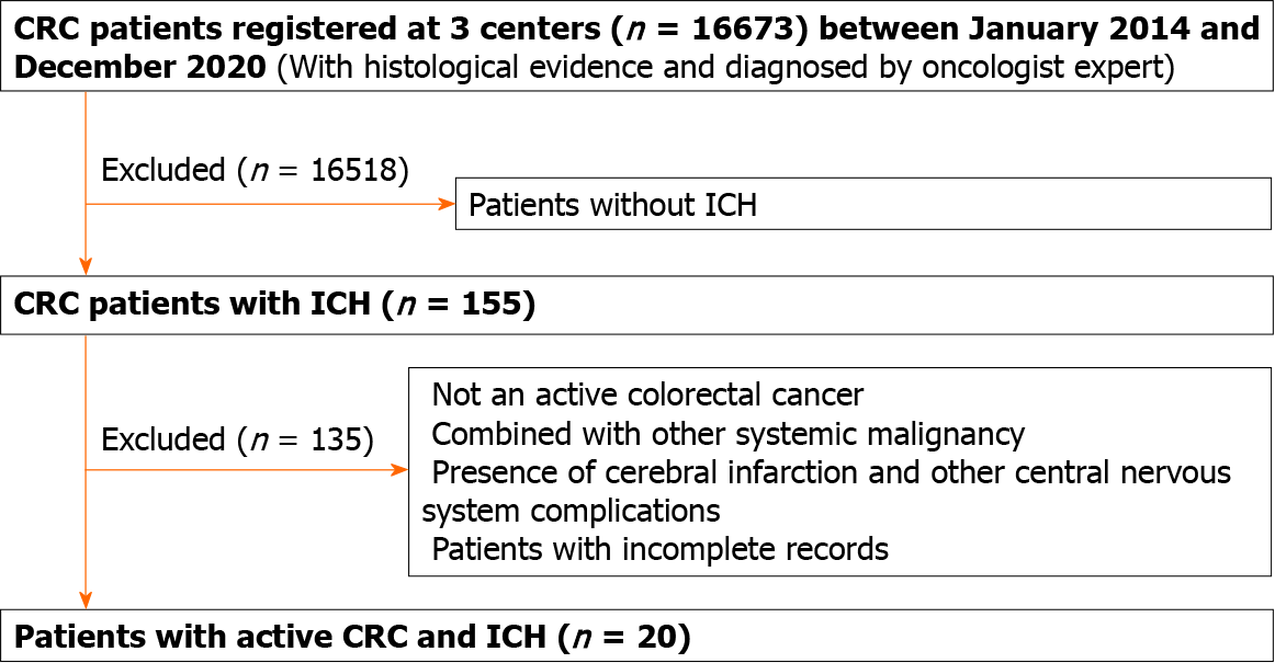
Figure 1 Patient enrollment flowchart.
CRC: Colorectal cancer; ICH: Intracerebral hemorrhage.
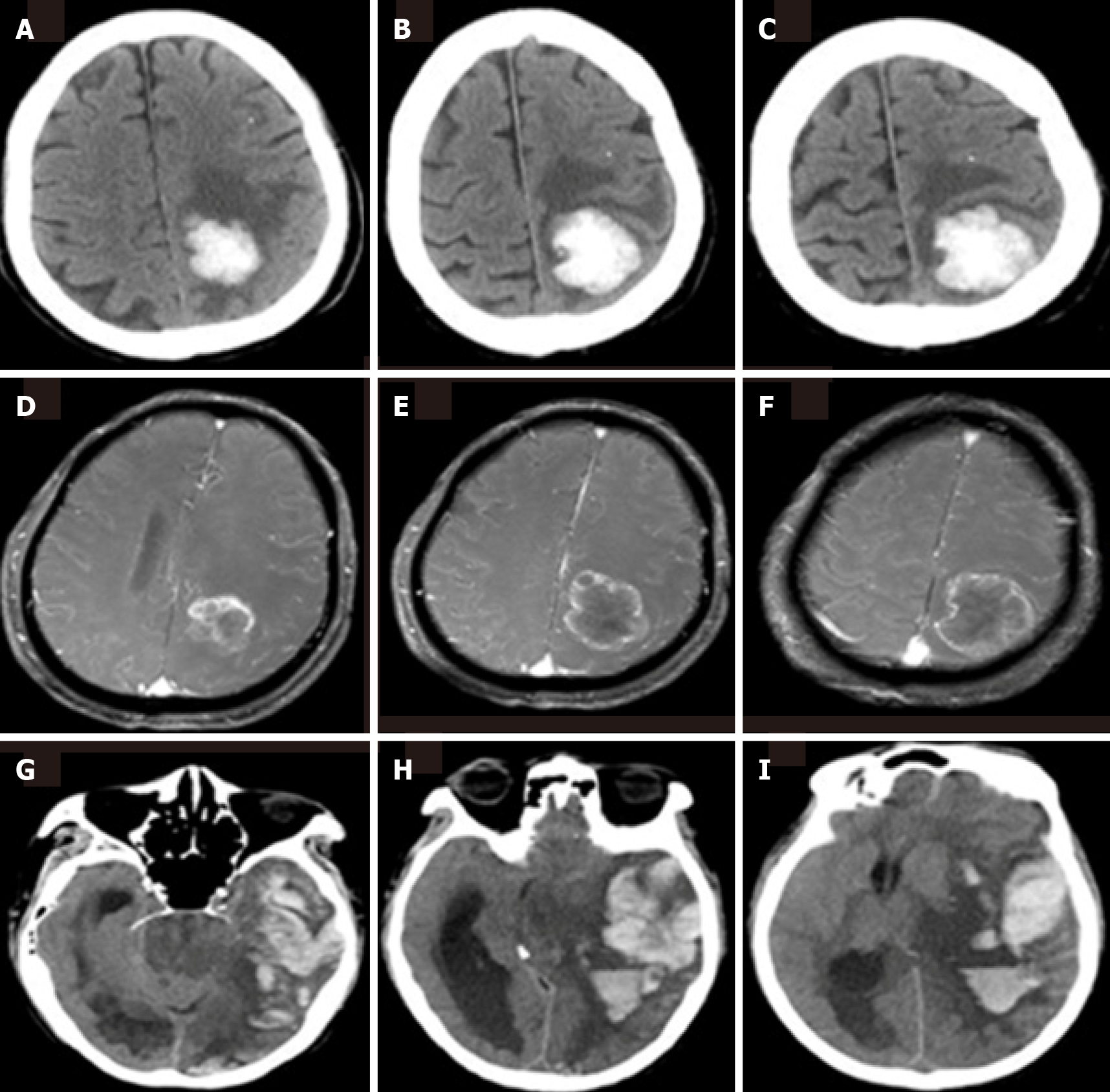
Figure 2 Neuroimaging findings.
A-F: Neuroimages of a 76-year-old patient with active colorectal cancer (CRC). Brain computed tomography axial views, showing a hemorrhagic lesion in the left parietal lobe (A-C); brain enhanced magnetic resonance views, showing a metastatic tumor in the same location (D-F); G-I: Neuroimages of a 65-year-old patient with active CRC and disseminated intravascular coagulation, showing a massive hemorrhage in the left temporal and parietal lobe.
- Citation: Deng XH, Li J, Chen SJ, Xie YJ, Zhang J, Cen GY, Song YT, Liang ZJ. Clinical features of intracerebral hemorrhage in patients with colorectal cancer and its underlying pathogenesis. World J Gastrointest Oncol 2021; 13(12): 2180-2189
- URL: https://www.wjgnet.com/1948-5204/full/v13/i12/2180.htm
- DOI: https://dx.doi.org/10.4251/wjgo.v13.i12.2180


