Copyright
©The Author(s) 2021.
World J Gastrointest Oncol. Dec 15, 2021; 13(12): 1863-1879
Published online Dec 15, 2021. doi: 10.4251/wjgo.v13.i12.1863
Published online Dec 15, 2021. doi: 10.4251/wjgo.v13.i12.1863
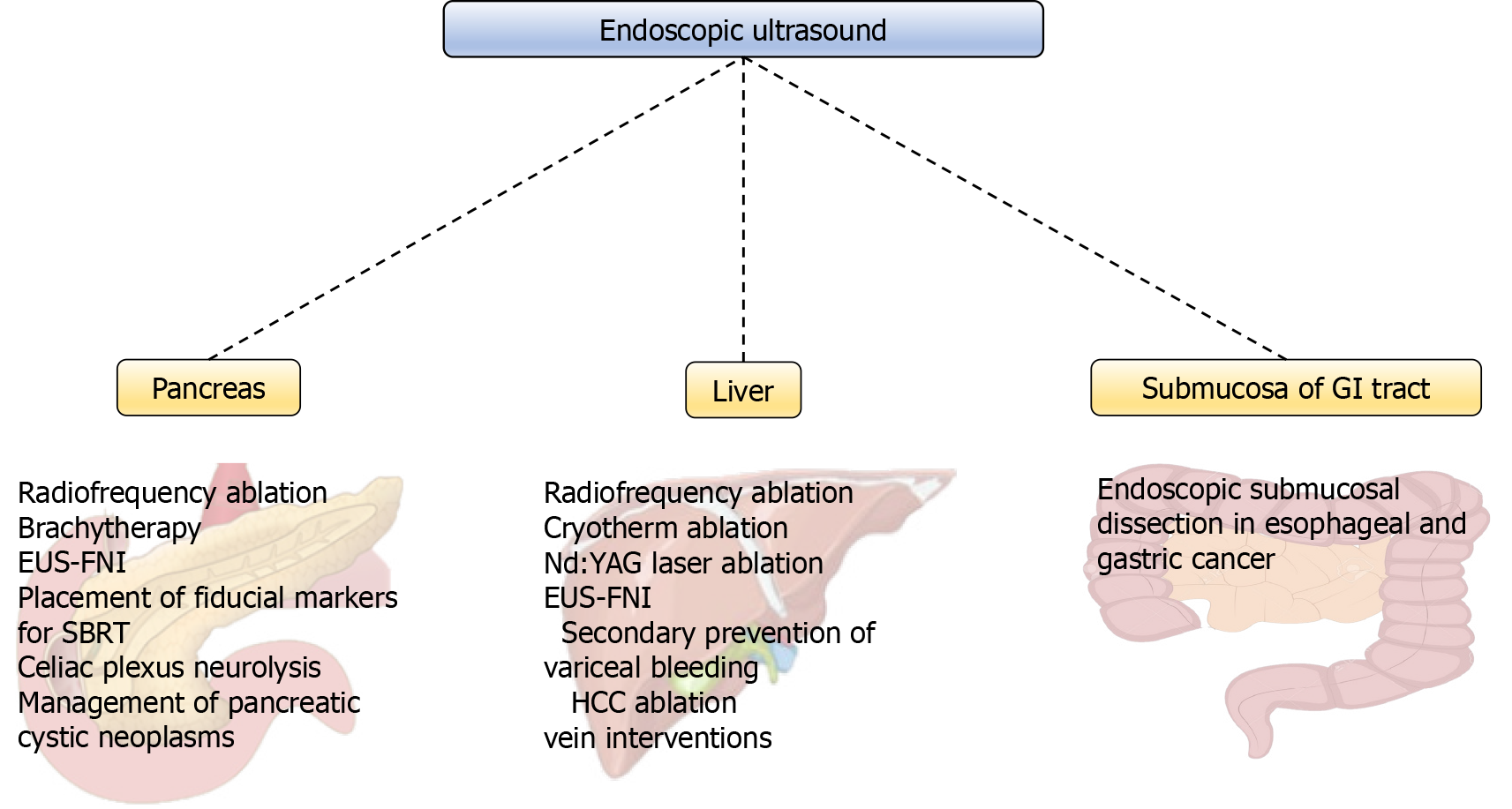
Figure 1 Overview of endoscopic ultrasound-guided methods.
EUS-FNI: Endoscopic ultrasound-fine needle injection; HCC: Hepatocellular carcinoma; GI: Gastrointestinal; Nd:YAG: Neodymium-doped yttrium aluminum garnet; SBRT: Stereotactic body radiotherapy.
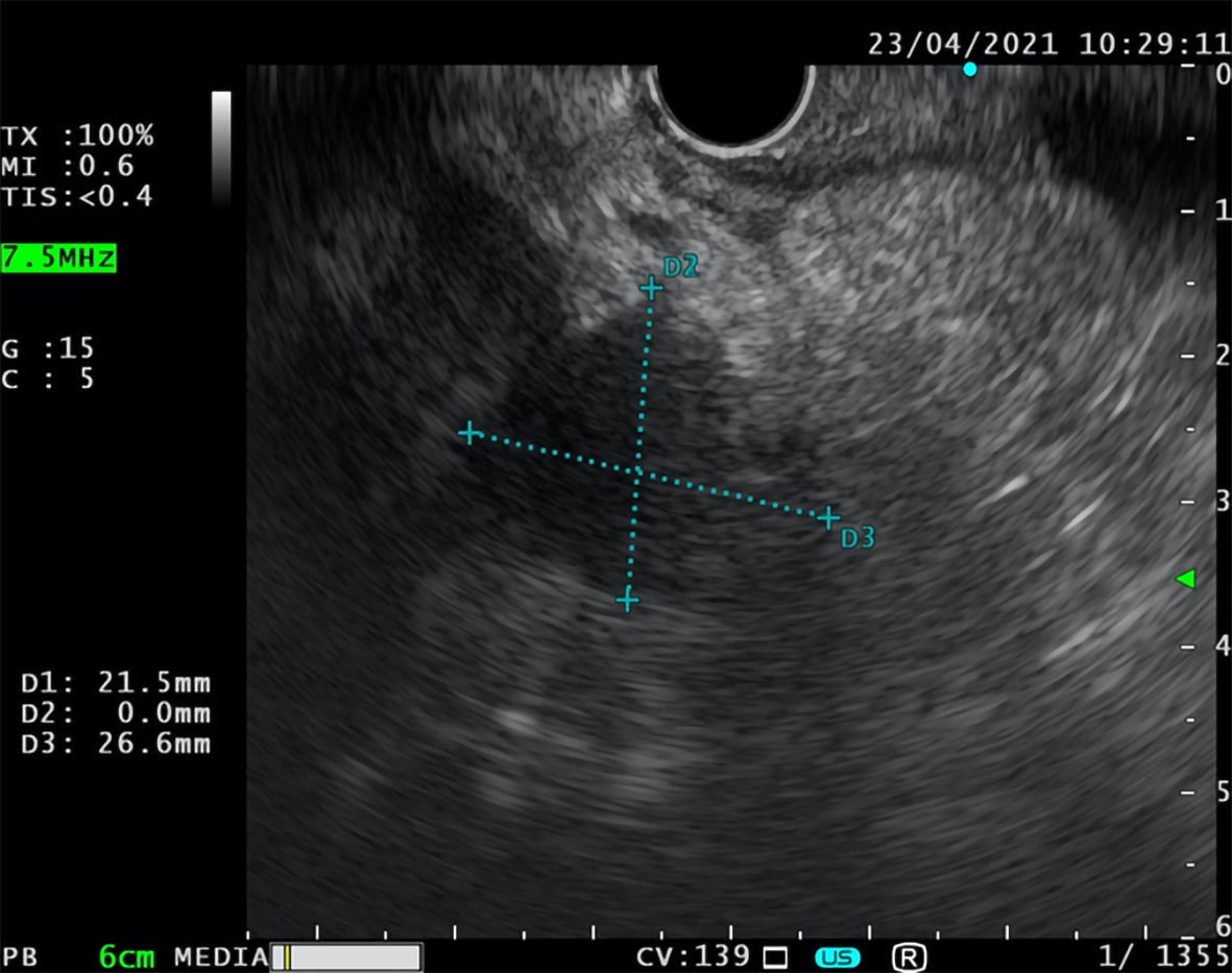
Figure 2 Endoscopic ultrasound-fine needle aspiration.
Fine needle aspiration of inhomogeneous oval lesion located on the border between head and corpus of the pancreas (26.6 mm × 21.5 mm).
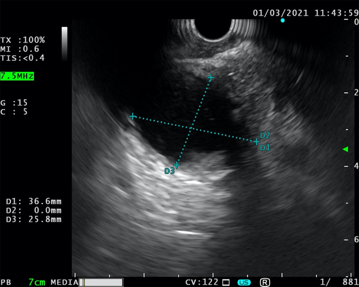
Figure 3 Unsuccessful endoscopic ultrasound-fine needle aspiration.
Unsuccessful attempt of fine needle aspiration of the cystic lesion with thick, calcified border (37 mm × 26 mm) located in the head of the pancreas.
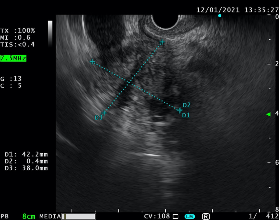
Figure 4 Endoscopic ultrasound-fine needle biopsy.
Fine needle biopsy of the focal lesion in the pancreatic head (42 mm × 38 mm).
- Citation: Bratanic A, Bozic D, Mestrovic A, Martinovic D, Kumric M, Ticinovic Kurir T, Bozic J. Role of endoscopic ultrasound in anticancer therapy: Current evidence and future perspectives. World J Gastrointest Oncol 2021; 13(12): 1863-1879
- URL: https://www.wjgnet.com/1948-5204/full/v13/i12/1863.htm
- DOI: https://dx.doi.org/10.4251/wjgo.v13.i12.1863


