Copyright
©The Author(s) 2020.
World J Gastrointest Oncol. Jul 15, 2020; 12(7): 719-731
Published online Jul 15, 2020. doi: 10.4251/wjgo.v12.i7.719
Published online Jul 15, 2020. doi: 10.4251/wjgo.v12.i7.719
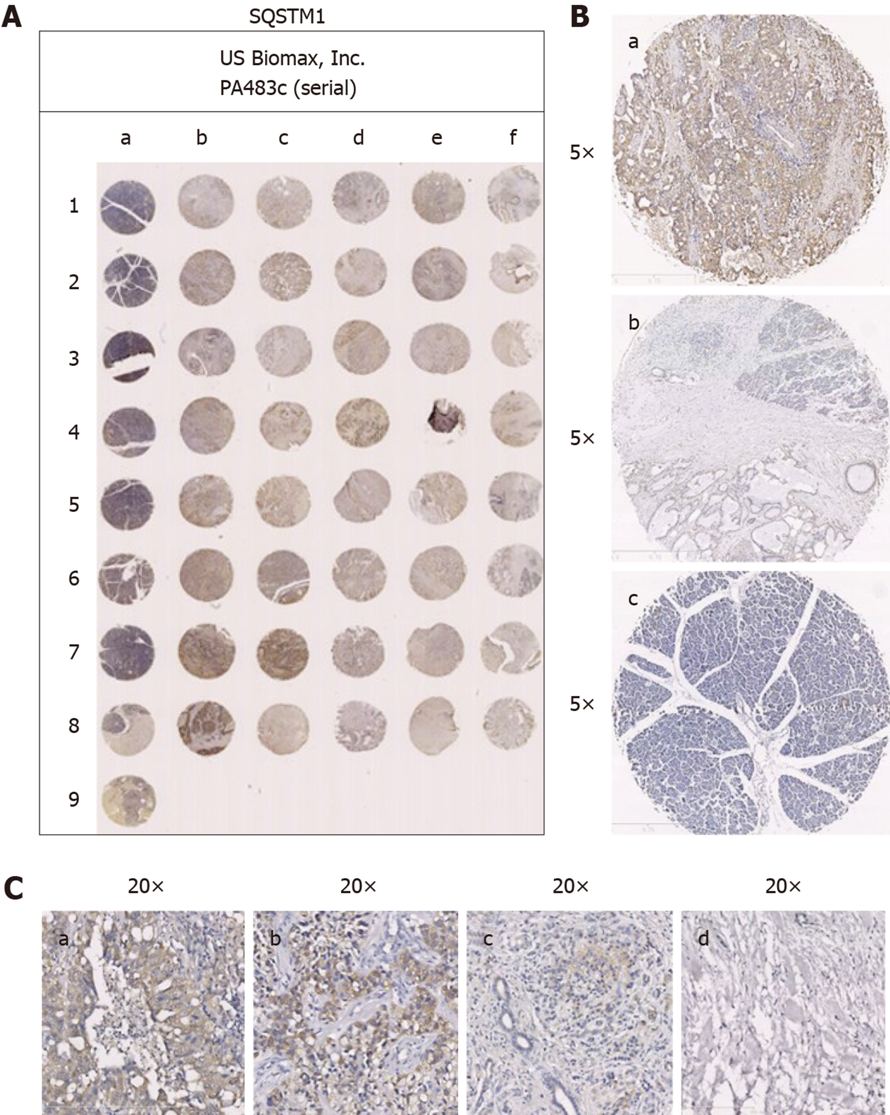
Figure 1 Photomicrographs of immunohistochemical staining for SQSTM1 (P62) in human samples of pancreatic normal tissues and pancreatic carcinoma tissues.
A: Expression of P62 in pancreatic normal tissues and pancreatic carcinoma tissues; B: (a) Strongly positive expression of P62 in pancreatic carcinoma tissue (5 ×); (b) Weakly positive expression of P62 in tumor invading edge sample (5 ×); and (c) Negative expression of P62 in pancreatic normal tissue (5 ×); C: (a) Strongly positive expression of P62 in pancreatic carcinoma tissue (20 ×); (b) Positive expression of P62 in pancreatic carcinoma tissue (20 ×); (c) Weakly positive expression of P62 in pancreatic carcinoma tissue (20 ×); and (d) Negative expression of P62 in pancreatic carcinoma tissue (20 ×).
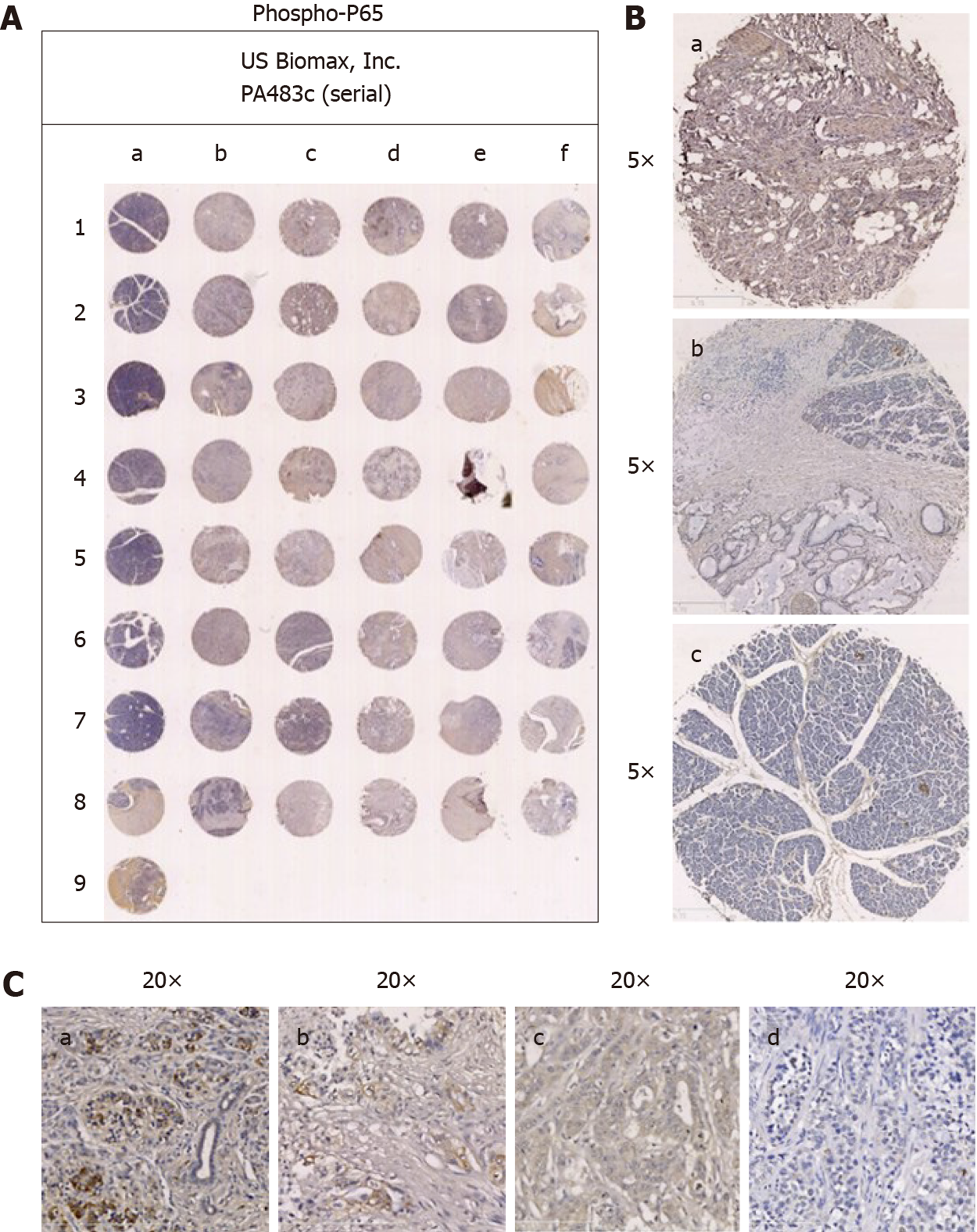
Figure 2 Photomicrographs of immunohistochemical staining for phospho-P65 in human samples of pancreatic normal tissues and pancreatic carcinoma tissues.
A: Expression of phospho-P65 in pancreatic normal tissues and pancreatic carcinoma tissues; B: (a) Strongly positive expression of phospho-P65 in pancreatic carcinoma tissue (5 ×); (b) Weakly positive expression of phospho-P65 in tumor invading edge sample (5 ×); and (c) Negative expression of phospho-P65 in pancreatic normal tissue (5 ×); C: (a) Strongly positive expression of phospho-P65 in pancreatic carcinoma tissue (20 ×); (b) Positive expression of phospho-P65 in pancreatic carcinoma tissue (20 ×); (c) Weakly positive expression of phospho-P65 in pancreatic carcinoma tissue (20 ×); and (d) Negative expression of phospho-P65 in pancreatic carcinoma tissue (20 ×).
- Citation: Zhang ZY, Guo S, Zhao R, Ji ZP, Zhuang ZN. Clinical significance of SQSTM1/P62 and nuclear factor-κB expression in pancreatic carcinoma. World J Gastrointest Oncol 2020; 12(7): 719-731
- URL: https://www.wjgnet.com/1948-5204/full/v12/i7/719.htm
- DOI: https://dx.doi.org/10.4251/wjgo.v12.i7.719


