Copyright
©The Author(s) 2018.
World J Gastrointest Oncol. Oct 15, 2018; 10(10): 317-327
Published online Oct 15, 2018. doi: 10.4251/wjgo.v10.i10.317
Published online Oct 15, 2018. doi: 10.4251/wjgo.v10.i10.317
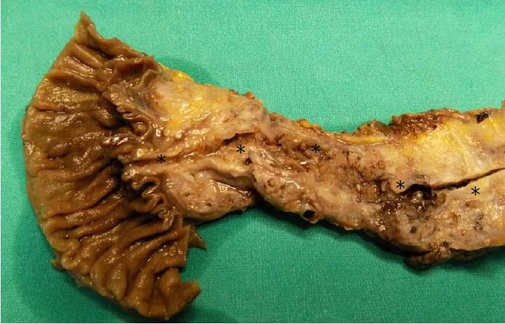
Figure 1 Extensive involvement of the pancreas by an intraductal papillary mucinous neoplasm.
This neoplasm involves almost all the pancreatic ductal tree. The asterisks indicate Wirsung's duct along its course.
Figure 2 Pancreatic intraepithelial neoplasm precursor lesions.
A: High-grade pancreatic intraepithelial neoplasm (PanIN); B: Low-grade PanIN. Original magnification: × 10. Black arrows indicate ducts involved by PanINs.
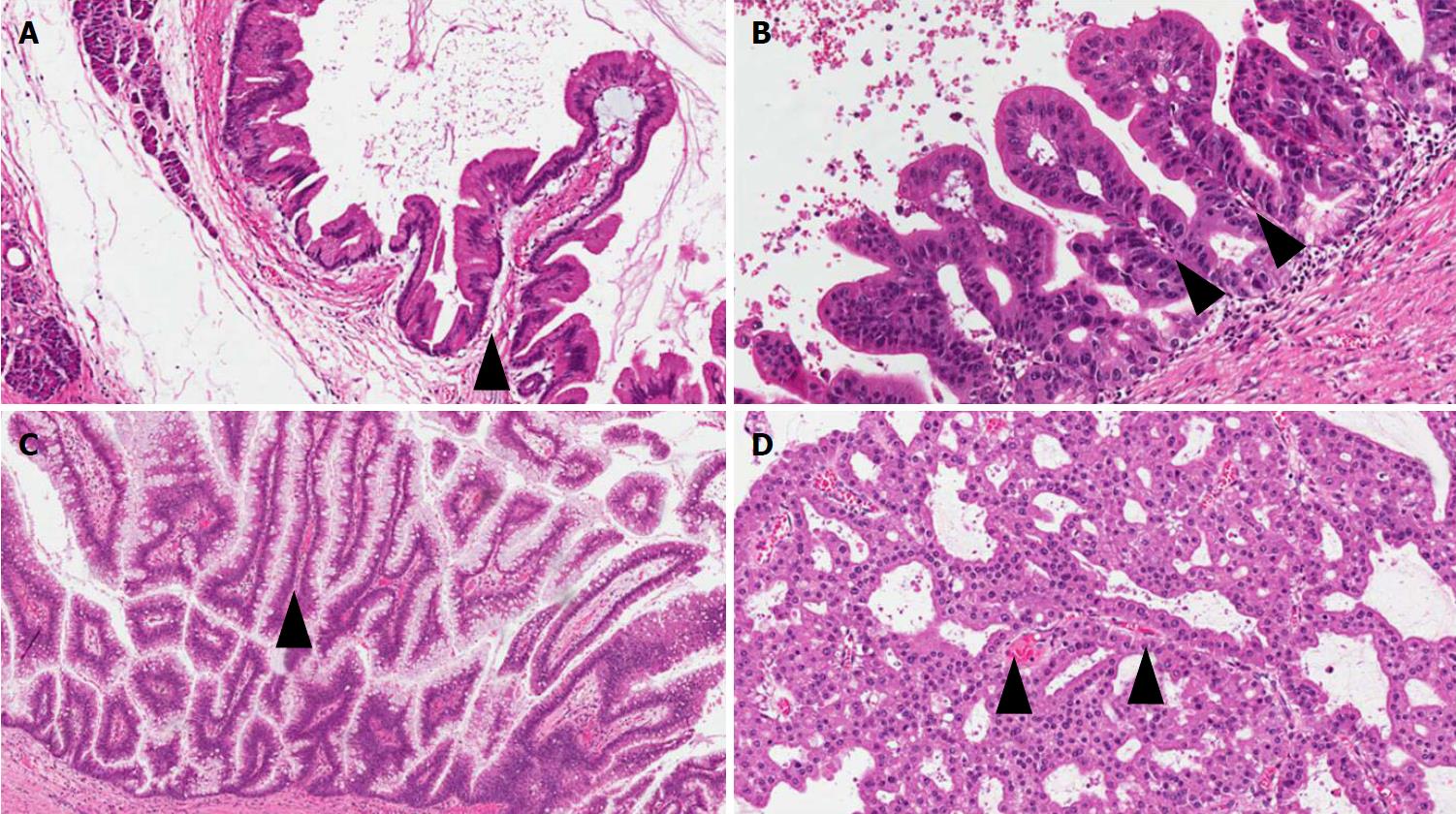
Figure 3 The four different types of intraductal papillary mucinous neoplasm.
A: Gastric; B: Pancreatobiliary; C: Intestinal; D: Oncocytic. Original magnification: A and C: × 10; B and D: × 20. Black arrowheads indicate the fibro-vascular axis of the papillary structures.
Figure 4 Colloid carcinoma with perineural invasion (A, black arrow) and nodal metastasis (B: low magnification; C: higher magnification of the same metastasis).
Original magnification: A: × 10; B: × 4; C: × 20.
Figure 5 Mucinous cystic neoplasm precursor lesions.
A: Low-grade mucinous cystic neoplasm (MCN); B: High-grade MCN. The black arrow indicates the ovarian-like stroma, a typical component of this type of lesion. Original magnification: × 10.
Figure 6 Intratubular papillary neoplasm precursor lesions.
A: Low magnification showing an extensive intraductal growth; B: Higher magnification. Original magnification A: × 1; B: × 4.
- Citation: Riva G, Pea A, Pilati C, Fiadone G, Lawlor RT, Scarpa A, Luchini C. Histo-molecular oncogenesis of pancreatic cancer: From precancerous lesions to invasive ductal adenocarcinoma. World J Gastrointest Oncol 2018; 10(10): 317-327
- URL: https://www.wjgnet.com/1948-5204/full/v10/i10/317.htm
- DOI: https://dx.doi.org/10.4251/wjgo.v10.i10.317


