Published online Oct 25, 2015. doi: 10.4253/wjge.v7.i15.1186
Peer-review started: June 27, 2015
First decision: July 10, 2015
Revised: July 26, 2015
Accepted: September 13, 2015
Article in press: September 21, 2015
Published online: October 25, 2015
Processing time: 118 Days and 18.2 Hours
AIM: To demonstrate the feasibility and reproducibility of a pure natural orifice transluminal endoscopic surgery (NOTES) gastrojejunostomy using holing followed by interrupted suture technique using a single endoloop matched with a pair of clips in a non-survival porcine model.
METHODS: NOTES gastrojejunostomy was performed on three female domestic pigs as follows: Gastrostomy, selection and retrieval of a free-floating loop of the small bowel into the stomach pouch, hold and exposure of the loop in the gastric cavity using a submucosal inflation technique, execution of a gastro-jejunal mucosal-seromuscular layer approximation using holing followed by interrupted suture technique with endoloop/clips, and full-thickness incision of the loop with a Dual knife.
RESULTS: Pure NOTES side-to-side gastrojejunostomy was successfully performed in all three animals. No leakage was identified via methylene blue evaluation following surgery.
CONCLUSION: This novel technique for preforming a gastrointestinal anastomosis exclusively by NOTES is technically feasible and reproducible in an animal model but warrants further improvement.
Core tip: A pure natural orifice transluminal endoscopic surgery gastrojejunostomy procedure may be successfully performed in a non-survival porcine model using holing followed by interrupted suture technique using one endoloop matched with a pair of clips, without the need of any additional devices.
- Citation: Chen SY, Shi H, Jiang SJ, Wang YG, Lin K, Xie ZF, Liu XJ. Transgastric endoscopic gastrojejunostomy using holing followed by interrupted suture technique in a porcine model. World J Gastrointest Endosc 2015; 7(15): 1186-1190
- URL: https://www.wjgnet.com/1948-5190/full/v7/i15/1186.htm
- DOI: https://dx.doi.org/10.4253/wjge.v7.i15.1186
Gastro-jejunal side-to-side anastomosis is clinically designed for palliation of malignant gastric outlet obstruction (GOO)[1], performed primarily via open[2] and laparoscopic surgery[3]. Natural orifice transluminal endoscopic surgery (NOTES) may represent an alternative for the execution of gastro-jejunostomy procedures[4-10] due to less invasiveness and postoperative pain compared with the above-mentioned two procedures. To date, dozens of successful gastric bypass procedures by pure or hybrid NOTES have been reported, however, these methods are associated with some limitations, including being time-consuming, technically demanding and requiring specialized suturing devices.
Our experimental study aimed to demonstrate the feasibility and reproducibility of a pure NOTES gastrojejunostomy procedure using holing[11] followed by interrupted suture technique using a single endoloop matched with a pair of clips[12] in a non-survival porcine model.
Our study involved three healthy female domestic pigs weighing between 15 and 20 kg. All animals were fasted for 24 h prior to surgery. Induction of anesthesia was achieved via an intramuscular injection of 100 mg ketamine, 10 mg droperidol and 1 mg atropine, and anesthesia was maintained by intravenous drip of propofol at a rate of 10 mL/h with endotracheal intubation. Heart rate and oxygen saturation were monitored during the operation. Animals were kept in a supine position to allow for the best access and optimal peritoneal exploration. This study was approved by the Institutional Animal Use and Care Committee of Fujian Provincial Tumor Hospital, Teaching Hospital of Fujian Medical University, Fuzhou, China.
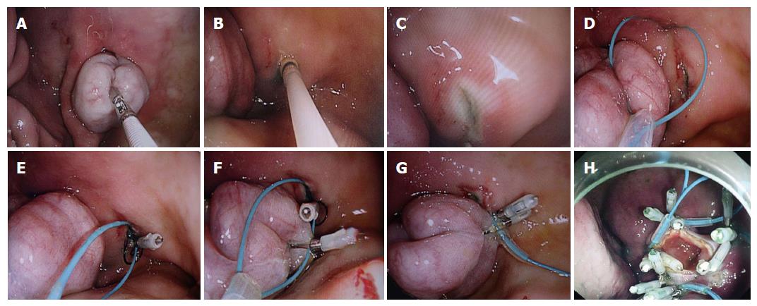
Gastrostomy: A small incision was created in the horizontal portion of the anterior pre-antral zone, which was determined via finger indentations of the abdominal wall, away from the small and large curvature, using a Dual knife (KD650L Olympus), followed by dilation using an 18-mm CRE balloon. The dual-channel therapeutic endoscope (GIF2TQ260M, Olympus Tokyo Japan) was subsequently advanced into the peritoneal cavity through the gastrostomy site.
Selection and retrieval of a free-floating loop of the small bowel into the stomach pouch: Loop selection was guided by loop proximity to the gastrostomy site to minimize the risk of tension and possible ischemia. An appropriate segment of the upper small intestine on its anti-mesenteric side was grasped by an endoscopic alligator forceps (FQ-46L-1, Olympus) through one channel of the endoscope and dragged through the incision into the stomach for the intra-gastric anastomosis, taking care not to include the mesenteric vascular supply to avoid unexpected incarceration.
Hold and exposure of the loop in the gastric cavity via submucosal inflation: An endoscopic injector (NM-400L-0423 Olympus) was passed through the other channel of the endoscope. Five to ten mL mL of saline solution mixed with 0.1 mL of 2% methylene blue was immediately injected into the submucosal layer circumferentially along the periphery of the gastrostomy site. Submucosal inflation temporarily decreased the size of the orifice of the gastrostomy to prevent the loop from falling back to the peritoneal cavity.
Execution of a gastro-jejunal mucosal-seromuscular layer approximation using holing followed by interrupted suture technique with endoloop/clips: First, a total of five to seven holes were made circumferentially along the periphery of the gastrostomy by using the Dual knife. An endoloop followed by an endoclip delivery system was inserted into the gastric cavity through the double-channel endoscope and placed at the side of one hole. One prong of the clip was then inserted in the hole of the stomach wall and clipped to anchor the endoloop. The second clip was used to anchor the same endoloop to the serosal surface of the small intestine. The gastric mucosal layer and the intestinal serosal layer were approximated by tightening of the endoloop. Briefly, gastro-jejunal mucosal-seromuscular layer anastomosis was created in pairs through the mucosa of the stomach and the serosa of small intestine to join the tissues based on the cooperation between one loop and a pair of clips. Five to seven pairs of interrupted sutures were placed to secure the anastomosis.
Full-thickness incision of the loop with the Dual knife: Jejunal loop incision was made longitudinally on its anti-mesenteric aspect to turn the inside mucosa out.
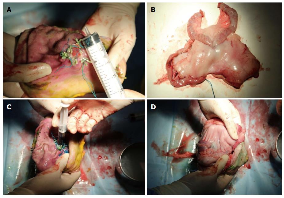
Euthanasia was performed immediately after the procedure. Necropsy results including injuries to adjacent organs, vascular bleeding, anastomotic patency and leakage evaluation were recorded.
Detailed data of pure NOTES side-to-side gastrojejunostomy performed on the three animals are shown in Table 1 and Figure 1. The procedure was technically successful in all cases. The duration of the procedure ranged from 1.0 to 1.5 h. Minor bleeding occurred from the right gastroepiploic artery during gastrostomy in 2 pigs and treated efficiently with the endoscopic hemostatic forceps (FD-410LR, Olympus) (80 W/soft-coagulation). On the postmortem examination, the immediate patency of the anastomosis was satisfactory, and no evidence of anastomotic leakage was identified via methylene blue evaluation[13] (Figure 2).
| Observation parameters | Pig 1 | Pig 2 | Pig 3 |
| Time required to enter the peritoneal cavity and pull the loop intro the stomach (min) | 35 | 19 | 18 |
| Time required to suture the anastomosis (min) | 58 | 44 | 47 |
| Number of the stitched pairs | 5 | 6 | 7 |
| Complications | |||
| Minor bleeding | + | + | - |
| Anastomotic leak | - | - | - |
The advent of NOTES has made a minimally invasive endoscopic technique possible for creation of gastrojejunal anastomosis, being faced with opportunities and challenges at the same time.
Previous studies[4,7,8-10] have reported three full-thickness suturing methods summarized as the small intestine being pulled into the stomach lumen and then sutured to the stomach wall using newly designed endoscopic suturing devices as follows: (1) a prototype endoscopic suturing device (Eagle Claw; Olympus)[7]; (2) a prototype “T-tag” suture system (BraceBar; Olympus)[4,9,10]; and (3) an EndoGIA stapler (Covidien)[8]. Here we reported for the first time, the use of interrupted suture technique using one endoloop matched with a pair of clips in a non-survival porcine model. This new technique resembles T-tag suture system, one of the aforementioned three methods, with its own unique characteristics. First, submucosal saline solution injection around the gastrostomy site made a slight cushion that prevented unexpected perforation by electric knives and allowed space to create holes deep enough to insert the prongs of the clip and to facilitate the subsequent secure clipping. Furthermore, submucosal inflation temporarily decreased the size of the orifice, and the smaller orifice allowed us to manipulate the loop of the small intestine in place more easily. Second, creating several holes at the edge of gastrostomy provided strong anchoring points for one prong of a clip in order to avoid clip slippage during grasping gastric thick mucosal surface. Third, interrupted suture method using one endoloop matched with a pair of clips was derived from the principle of “sewing” using a pair of T-tags with a single puncture needle, which may be done successfully by using only endoscopes and common endoscopic accessories, without the need of any extra devices.
In particular, if the small intestine was inadvertently dropped during the procedure, no leakage of small bowel contents occurred because the bowel wall was not incised until the anastomosis was complete.
Our pilot study had several limitations, however. First, endoscopic selection of an appropriate loop of the small bowel and secure fixation of the small bowel to the gastric wall without intra-peritoneal manipulation remains challenging[14]. In our current study, the appropriate portion of the small bowel was identified based on its proximity to the gastrostomy site and the left upper abdominal anatomical landmarks such as the spleen. For clinical studies, EUS guidance could be used to direct the targeted jejunal segment near the ligament of Treitz in non-altered anatomy patients[1,15]. Second, both of the endoscopic endoloop/clips utilized in our study, as well as the sewing devices (such as T-bar sutures) predominantly approximate the mucosa, and the reliability and durability of the anastomosis under gastric pressure should be estimated in the porcine model of GOO. Ryou et al[16] demonstrated that gastric mucosal closure with endoscopic clips may result in significant air and fluid leakage via the line of clips, however, this was not observed in our study.
In conclusion, this novel technique of performing gastrointestinal anastomosis exclusively by NOTES is technically feasible and reproducible in an animal model, although further improvement is warranted.
Gastro-jejunal side-to-side anastomosis is clinically designed for palliation of malignant gastric outlet obstruction (GOO), mostly performed via open and laparoscopic surgery. Natural orifice transluminal endoscopic surgery may represent an alternative method of performing gastro-jejunostomy procedures due to its less invasiveness and lower incidence of postoperative pain compared with the above-mentioned two methods.
To date, dozens of successful gastric bypass procedures via either pure or hybrid natural orifice transluminal endoscopic surgery (NOTES) have been described, however, these methods are associated with some limitations, as they are time-consuming, technically demanding and require specialized suturing devices.
A pure NOTES gastrojejunostomy procedure may be successfully performed in a non-survival porcine model using holing followed by interrupted suture technique using one endoloop matched with a pair of clips, without the need of any additional devices.
This study demonstrates the potential application of pure NOTES gastrojejunostomy using holing followed by interrupted suture technique using one endoloop matched with a pair of clips for palliation of malignant GOO.
A NOTES gastrojejunostomy using interrupted suture technique with a single endoloop matched with a pair of clips resembles the technical principle of T-tag suture system, without the need of any additional devices.
The advent of NOTES has made a minimally invasive endoscopic technique possible for creation of gastrojejunal anastomosis, being faced with opportunities and challenges at the same time. A pure NOTES gastrojejunostomy procedure may be successfully performed in a non-survival porcine model using holing followed by interrupted suture technique using one endoloop matched with a pair of clips.
P- Reviewer: Tomizawa M S- Editor: Ma YJ L- Editor: Wang TQ E- Editor: Jiao XK
| 1. | Khashab MA, Baron TH, Binmoeller KF, Itoi T. EUS-guided gastroenterostomy: a new promising technique in evolution. Gastrointest Endosc. 2015;81:1234-1236. [RCA] [PubMed] [DOI] [Full Text] [Cited by in Crossref: 30] [Cited by in RCA: 39] [Article Influence: 3.9] [Reference Citation Analysis (35)] |
| 2. | Chandrasegaram MD, Eslick GD, Mansfield CO, Liem H, Richardson M, Ahmed S, Cox MR. Endoscopic stenting versus operative gastrojejunostomy for malignant gastric outlet obstruction. Surg Endosc. 2012;26:323-329. [RCA] [PubMed] [DOI] [Full Text] [Cited by in Crossref: 71] [Cited by in RCA: 67] [Article Influence: 5.2] [Reference Citation Analysis (0)] |
| 3. | Mehta S, Hindmarsh A, Cheong E, Cockburn J, Saada J, Tighe R, Lewis MP, Rhodes M. Prospective randomized trial of laparoscopic gastrojejunostomy versus duodenal stenting for malignant gastric outflow obstruction. Surg Endosc. 2006;20:239-242. [PubMed] |
| 4. | Bergström M, Ikeda K, Swain P, Park PO. Transgastric anastomosis by using flexible endoscopy in a porcine model (with video). Gastrointest Endosc. 2006;63:307-312. [PubMed] |
| 5. | Yi SW, Chung MJ, Jo JH, Lee KJ, Park JY, Bang S, Park SW, Song SY. Gastrojejunostomy by pure natural orifice transluminal endoscopic surgery using a newly designed anastomosing metal stent in a porcine model. Surg Endosc. 2014;28:1439-1446. [RCA] [PubMed] [DOI] [Full Text] [Cited by in Crossref: 8] [Cited by in RCA: 8] [Article Influence: 0.7] [Reference Citation Analysis (0)] |
| 6. | Barthet M, Binmoeller KF, Vanbiervliet G, Gonzalez JM, Baron TH, Berdah S. Natural orifice transluminal endoscopic surgery gastroenterostomy with a biflanged lumen-apposing stent: first clinical experience (with videos). Gastrointest Endosc. 2015;81:215-218. [PubMed] |
| 7. | Kantsevoy SV, Jagannath SB, Niiyama H, Chung SS, Cotton PB, Gostout CJ, Hawes RH, Pasricha PJ, Magee CA, Vaughn CA. Endoscopic gastrojejunostomy with survival in a porcine model. Gastrointest Endosc. 2005;62:287-292. [PubMed] |
| 8. | Chiu PW, Wai Ng EK, Teoh AY, Lam CC, Lau JY, Sung JJ. Transgastric endoluminal gastrojejunostomy: technical development from bench to animal study (with video). Gastrointest Endosc. 2010;71:390-393. [RCA] [PubMed] [DOI] [Full Text] [Cited by in Crossref: 22] [Cited by in RCA: 24] [Article Influence: 1.6] [Reference Citation Analysis (0)] |
| 9. | Vanbiervliet G, Gonzalez JM, Bonin EA, Garnier E, Giusiano S, Saint Paul MC, Berdah S, Barthet M. Gastrojejunal Anastomosis Exclusively Using the “NOTES” Technique in Live Pigs: A Feasibility and Reliability Study. Surg Innov. 2014;21:409-418. [PubMed] |
| 10. | Song TJ, Seo DW, Kim SH, Park do H, Lee SS, Lee SK, Kim MH. Endoscopic gastrojejunostomy with a natural orifice transluminal endoscopic surgery technique. World J Gastroenterol. 2013;19:3447-3452. [RCA] [PubMed] [DOI] [Full Text] [Full Text (PDF)] [Cited by in CrossRef: 8] [Cited by in RCA: 9] [Article Influence: 0.8] [Reference Citation Analysis (0)] |
| 11. | Kim HH, Park SJ, Park MI, Moon W. Endoscopic hole and clipping technique: a novel technique for closing the wide orifice of a postoperative fistula. Gastrointest Endosc. 2012;75:1250-1252. [RCA] [PubMed] [DOI] [Full Text] [Cited by in Crossref: 3] [Cited by in RCA: 4] [Article Influence: 0.3] [Reference Citation Analysis (0)] |
| 12. | Zhang Y, Wang X, Xiong G, Qian Y, Wang H, Liu L, Miao L, Fan Z. Complete defect closure of gastric submucosal tumors with purse-string sutures. Surg Endosc. 2014;28:1844-1851. [RCA] [PubMed] [DOI] [Full Text] [Cited by in Crossref: 39] [Cited by in RCA: 53] [Article Influence: 4.8] [Reference Citation Analysis (0)] |
| 13. | Vegunta RK, Rawlings AL, Jeziorczak PM. Methylene blue: a simple marker for intraoperative detection of gastroduodenal perforations during laparoscopic pyloromyotomy. Surg Innov. 2010;17:11-13. [RCA] [PubMed] [DOI] [Full Text] [Cited by in RCA: 2] [Reference Citation Analysis (0)] |
| 14. | Luo H, Pan Y, Min L, Zhao L, Li J, Leung J, Xue L, Yin Z, Liu X, Liu Z. Transgastric endoscopic gastroenterostomy using a partially covered occluder: a canine feasibility study. Endoscopy. 2012;44:493-498. [RCA] [PubMed] [DOI] [Full Text] [Cited by in Crossref: 6] [Cited by in RCA: 9] [Article Influence: 0.7] [Reference Citation Analysis (0)] |
| 15. | Itoi T, Ishii K, Tanaka R, Umeda J, Tonozuka R. Current status and perspective of endoscopic ultrasonography-guided gastrojejunostomy: endoscopic ultrasonography-guided double-balloon-occluded gastrojejunostomy (with videos). J Hepatobiliary Pancreat Sci. 2015;22:3-11. [RCA] [PubMed] [DOI] [Full Text] [Cited by in Crossref: 37] [Cited by in RCA: 49] [Article Influence: 4.5] [Reference Citation Analysis (35)] |
| 16. | Ryou M, Pai RD, Sauer JS, Rattner DW, Thompson CC. Evaluating an optimal gastric closure method for transgastric surgery. Surg Endosc. 2007;21:677-680. [PubMed] |


