Published online Nov 16, 2014. doi: 10.4253/wjge.v6.i11.568
Revised: September 17, 2014
Accepted: October 1, 2014
Published online: November 16, 2014
Processing time: 197 Days and 18.5 Hours
This study reports a 69-year-old, obese, female patient presenting with a biliary leakage after laparoscopic cholecystectomy for cholelithiasis. Closure of the umbilical trocar site had been neglected during the laparoscopic cholecystectomy. Early, on postoperative day five, endoscopic retrograde cholangiopancreatography (ERCP) requirement after laparoscopic cholecystectomy resolved the biliary leakage problem but resulted with a more complicated clinical picture with an intestinal obstruction and severe abdominal pain. Computed tomography revealed a strangulated hernia from the umbilical trocar site. Increased abdominal pressure during ERCP had strained the weak umbilical trocar site. Emergency surgical intervention through the umbilicus revealed an ischemic small bowel segment which was treated with resection and anastomosis. This report demonstrates that negligence of trocar site closure can result in very early herniation, particularly if an endoscopic intervention is required in the early postoperative period.
Core tip: This report demonstrates that negligence of trocar site closure after laparoscopic surgery can result in very early trocar site herniation. This may occur particularly if an endoscopic intervention is required in the early postoperative period. Suturing the trocar site is not only important for the late trocar site herniations, it is imperative for early hernia problems as well.
- Citation: Sumer F, Kayaalp C, Yagci MA, Otan E, Kocaaslan H. Early endoscopic retrograde cholangiopancreatography after laparoscopic cholecystectomy can strain the occurrence of trocar site hernia. World J Gastrointest Endosc 2014; 6(11): 568-570
- URL: https://www.wjgnet.com/1948-5190/full/v6/i11/568.htm
- DOI: https://dx.doi.org/10.4253/wjge.v6.i11.568
Progress in minimally invasive procedures such as endoscopy or laparoscopy has resulted in revolutionary changes in clinical practice. Gastrointestinal endoscopy, particularly interventional procedures, have replaced some complicated surgical methods and improved patient care. On the other hand, the use of laparoscopy is increasing in most of the gastrointestinal surgical procedures. However, these minimal invasive procedures have themselves developed some very specific complications, particularly when they are combined. This study presents a case of strangulated perforated Richter’s hernia from the umbilical trocar site following laparoscopic cholecystectomy in the early postoperative period. This complication was accelerated by the requirement of early endoscopic retrograde cholangiopancreatography (ERCP) for the postoperative biliary leakage.
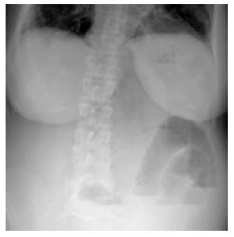
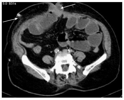
A 69-year-old female patient with a body mass index of 32 kg/m2 underwent laparoscopic cholecystectomy for cholelithiasis in a rural district hospital. On postoperative day one, abdominal drainage was 200 cc/d with bile content. The patient was referred to our center for diagnosis and treatment of the biliary leakage. The primary surgeon of the patient informed us that he identified the intact common bile duct during surgery but the procedure was difficult due to the inflammation of the gall bladder. Physical examination revealed no findings of peritonitis or evident abdominal distention. The laparoscopic cholecystectomy had been performed by conventional four trocars (two 11 mm and two 5 mm). Laboratory test results were within normal ranges. Abdominal ultrasound displayed a collection only 4 cm in diameter in the gall bladder bed. Because of the ongoing biliary leakage, an ERCP was performed on the 5th postoperative day. ERCP revealed a leakage from the cystic stump and a biliary 7F stent was placed. The procedure was completed uneventfully and the next day of ERCP the biliary leakage decreased immediately. However, an abrupt abdominal distention, pain and vomiting occurred after the ERCP procedure. Abdominal examination displayed tenderness, especially around the umbilicus. Obesity and previous surgery did not allow an objective examination of the umbilicus. Abdominal X-ray revealed air-fluid levels (Figure 1). Post-ERCP amylase value was 87 U/L (normal range: 36-128). Repeated abdominal ultrasound examinations showed that the previous collection at the gall bladder bed had disappeared. Her laboratory results were within normal limits during the two-day follow-up, but her symptoms (pain, vomiting and distention) persisted. Finally, a computed tomography on postoperative day 7, revealed the herniated intestine inside the umbilical trocar site (Figure 2). The patient underwent an emergency surgical treatment under general anesthesia. A laparotomy was performed containing the umbilical trocar site incision and an ileal anti-mesenteric loop was observed to be herniated. There was no previous fascia suturing at the umbilical trocar site. A small ileal segment of 3-4 cm in length was ischemic which consisted of a millimetric perforation. The rest of the abdominal exploration findings were normal. Following resection of the ischemic perforated ileal segment, a side-to-side intestinal anastomosis was performed. Postoperative course was uneventful and the patient was discharged on day eight of the second surgery.
The incidence of trocar site hernias in systematic reviews are generally reported as 1.7% (ranged 0.65%-2.80% or 0.3%-5.4%)[1,2], some studies reported a quite high incidence as 25.9%[3]. If we ignore the fact its incidence, they have the potential to cause serious complications[4]. Predisposing factors are using larger size trocars, leaving fascial defects open, midline positioned trocar sites, stretching the port sites for retrieving specimens, obesity, malnutrition, older age and surgical site infections[4]. Time period between the previous surgery and the diagnosis of trocar site hernia varies but reported as a mean 9.2 mo (ranged from 5 d to 3 years)[2]. In the very early postoperative period, they occur rarely and cause confusion in diagnosis if particularly accompanied with intestinal obstruction, strangulation or perforation. Diagnosis of hernia can be significantly difficult in obese patients. Infection and hematoma in the surgical site may also contribute to the under-diagnosis. Additionally, a co-existing intra-abdominal complication, which was a biliary leakage in our case, may lead to overlooking this relatively rare condition. Abdominal distention and pain can be considered to be related to the complications of the ERCP itself. Differential diagnosis needs careful clinical evaluation and radiological examinations.
Trocar site hernia after laparoscopic cholecystectomy usually include omentum or small bowel. In cases with presence of omentum in the sac, abdominal pain is generally mild[2]. Trocar site hernias following laparoscopic cholecystectomy are usually Richter hernias without strangulation, however, they rarely result in obstruction or perforation[2]. Computed tomography has several advantages for the evaluation of the abdominal wall over the other radiological methods, particularly in the patients with a high body mass index, mild clinical symptoms, hematoma or infection in the surgical site. Computed tomography is also beneficial for the differential diagnosis of the other postoperative complications. Persistent complaints and obscure physical examination findings confused us when making the diagnosis, but computed tomography revealed the umbilical trocar site hernia exactly.
Development of strangulation following upper gastrointestinal endoscopy or colonoscopy was reported in already known umbilical, diaphragmatic or Spigelian hernias[5]. However, there was no previous report considering the predisposing effect of endoscopic procedures (gastroscopy, colonoscopy or ERCP) during the early postoperative period on the development of trocar site herniation. The unknown hernia was diagnosed after the endoscopic procedure even though it had developed into a strangulated hernia. Early postoperative ERCP by increasing the intra-abdominal pressure promoted the trocar site hernia when the fascial defect was left open.
It was reported that following laparoscopic surgical procedures, 86.3% of the trocar site hernias were from incisions where trocars greater than 10 mm were used[6]. Although all the trocar defects greater than 10 mm should be closed as a rule, trocar site closures can sometimes be neglected with obese patients or after prolonged, difficult, devastating laparoscopic procedures. It is most likely for this reason that the primary surgeon of this case did not close the umbilical trocar site. Closure of trocar site fascial defects avoid trocar site hernias in the long-term follow-ups, however, they should be closed for the prevention of very early postoperative hernia complications.
As a conclusion, postoperative early endoscopic procedures may aggravate development of early herniation from trocar sites when fascial defects are left open.
The patient was referred to our center for diagnosis and treatment of the biliary leakage after laparoscopic cholecystectomy and a biliary 7F stent was placed by endoscopic retrograde cholangiopancreatography (ERCP) on the 5th postoperative day.
An abrupt abdominal distention, pain and vomiting occurred after the ERCP procedure.
Abdominal X-ray revealed air-fluid levels.
Post-ERCP amylase value was 87 U/L (normal range: 36-128).
Computed tomography on postoperative day 7, revealed the herniated intestine inside the umbilical trocar site. The patient underwent laparotomy for the diagnosis of umbilical trocar site herniation of the ileal anti-mesenteric loop. There was no previous fascia suturing at the umbilical trocar site.
A ileal segment of 3-4 cm in length was ischemic which consisted of a millimetric perforation.
Ischemic perforated ileal segment was resected.
There was no previous report considering the predisposing effect of endoscopic procedures (gastroscopy, colonoscopy or ERCP) during the early postoperative period on the development of trocar site herniation.
Suturing the trocar site is not only important for the late trocar site herniations, it is imperative for early hernia problems as well.
This case is interesting and relatively low in incidence.
P- Reviewer: Rabago L, Shim CS S- Editor: Ji FF L- Editor: A E- Editor: Zhang DN
| 1. | Mathews J. Incisional hernias after laparoscopic surgery. World J Laparoscopic Surg. 2010;1:13-17. |
| 3. | Comajuncosas J, Hermoso J, Gris P, Jimeno J, Orbeal R, Vallverdú H, López JL, Urgellés J, Estalella L, Parés D. Risk factors for umbilical trocar site incisional hernia in laparoscopic cholecystectomy: a prospective 3-year follow-up study. Am J Surg. 2014;207:1-6. |
| 4. | Comajuncosas J, Vallverdú H, Orbeal R, Parés D. [Trocar site incisional hernia in laparoscopic surgery]. Cir Esp. 2011;89:72-76. |
| 5. | Fulp SR, Gilliam JH. Beware of the incarcerated hernia. Gastrointest Endosc. 1990;36:318-319. |


