Published online Sep 16, 2010. doi: 10.4253/wjge.v2.i9.321
Revised: June 30, 2010
Accepted: July 7, 2010
Published online: September 16, 2010
Gallstone-induced ileus is a rare complication of cholelithiasis. Since localization of gallstones impacted in the small bowel, especially in the ileum, prevents access by conventional endoscopy in most cases, the mainstay of treatment remains surgical. Recent invention of double- and single-balloon enteroscopy has added much to the ability of imaging the small bowel and enables endoscopically directed therapy. Herein, for the first time, we report a successful endoscopic calculus removal via peroral single-balloon enteroscopy in an 81-year-old woman suffering from gallstone ileus of the ileum.
- Citation: Heinzow HS, Meister T, Wessling J, Domschke W, Ullerich H. Ileal gallstone obstruction: Single-balloon enteroscopic removal. World J Gastrointest Endosc 2010; 2(9): 321-324
- URL: https://www.wjgnet.com/1948-5190/full/v2/i9/321.htm
- DOI: https://dx.doi.org/10.4253/wjge.v2.i9.321
Although rarely reported, gallstone ileus is an important cause of mechanical bowel obstruction, resulting from impaction of a gallstone in the jejunum or ileum after being passed through a biliary-enteric fistula[1]. It was first described by Leon Bouveret in 1896. The diagnosis in mainly elderly patients who not infrequently have other significant medical conditions is often delayed since symptoms may be intermittent and investigations fail to identify the cause of obstruction[2]. Patients may present with nausea, vomiting and epigastric pain. Hematemesis can sometimes also occur due to erosion at the site of the biliary-enteric fistula[3]. Diagnostics usually include endoscopy, computed tomography (CT) and more recently magnetic resonance cholangiopancreatography. The diagnosis may be suggested by fulfilment of the Rigler triad: bowel obstruction, pneumobilia and ectopic gallstone. Since the reported mortality rate has is high at 12%, early diagnosis and treatment of gallstone-induced ileus remain crucial. So far, surgery has had the pivotal role in managing this condition. Enterolithotomy, cholecystectomy, and fistula division, with or without common bile duct exploration are the surgical options considered[1,4]. In high-risk patients, however, non-surgical treatment of gallstone ileus is desirable. Accordingly, successful electrohydraulic lithotripsy and extracorporeal shockwave lithotripsy (ESWL) of stones obstructing the jejunum, stomach, and colon has been reported in some cases[5-8]. Removal of gallstones from the upper small intestine via conventional endoscopy has also been described[9].
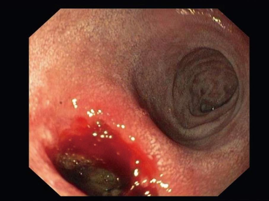
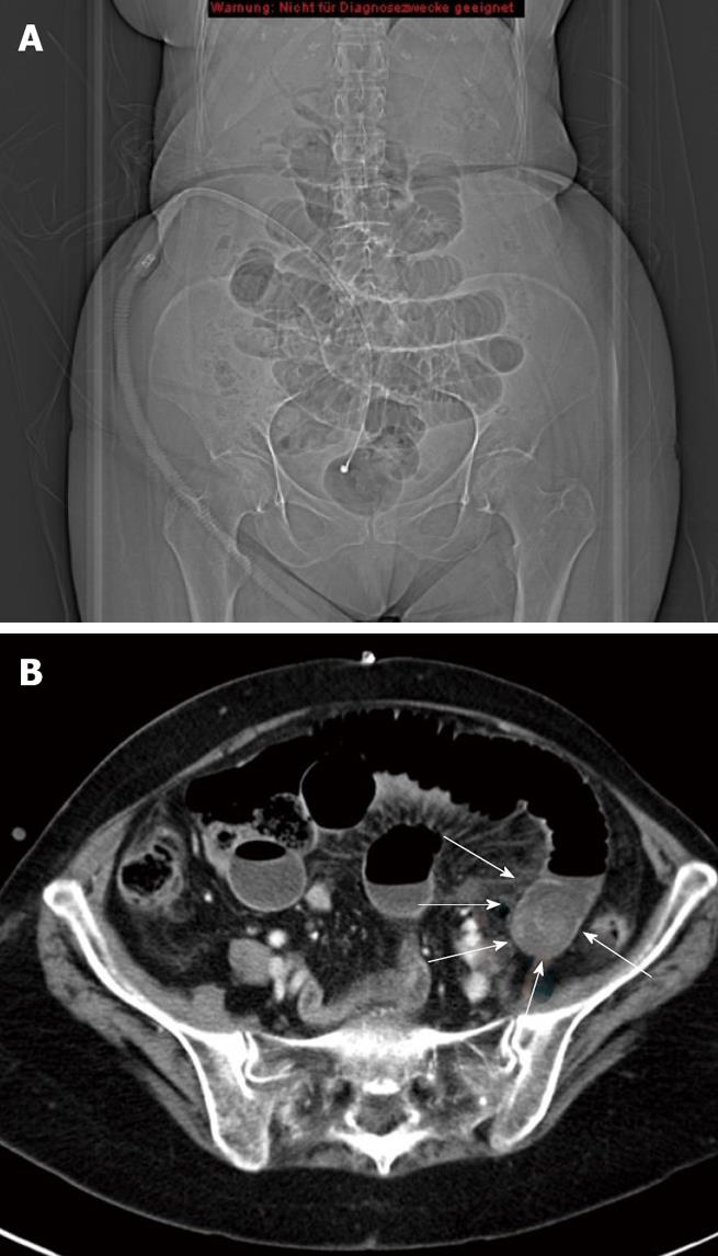
A febrile and slightly dehydrated 81-year-old woman was admitted due to a three day history of diffuse abdominal pain, nausea and hematemesis. On admission physical examination revealed mild tenderness of the upper abdominal quadrants and signs of subileus. There was no jaundice observed. Leukocytes and C-reactive protein were slightly elevated. Abdominal ultrasound showed pneumobilia. Upper gastrointestinal endoscopy revealed a deep ulcer of the duodenal bulb (Figure 1). Small bowel ileus was confirmed by plain abdominal X-ray and CT-scan showing the typical image of mechanical small bowel obstruction. Moreover, pneumobilia of the central biliary tract was observed(Figure 2A). Abdominal CT-scan (Figure 2B) also detected a fistulous structure extending from the duodenum to the gallbladder and, in addition, showed a calcified mass of 5 cm in diameter in the distal small bowel, thus establishing the diagnosis of bilioduodenal fistula and gallstone ileus of the ileum.
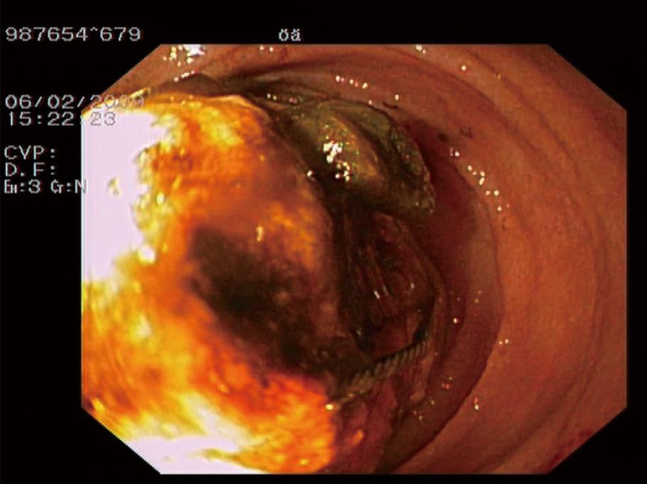
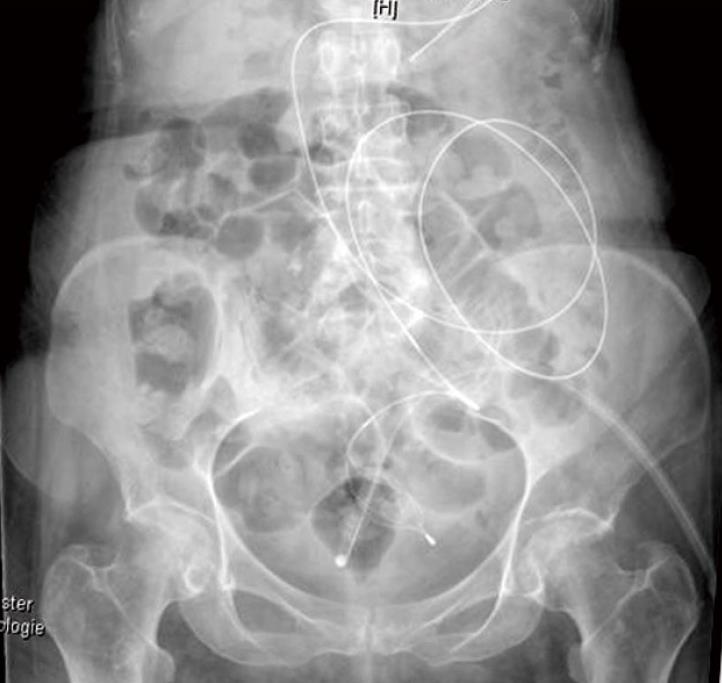
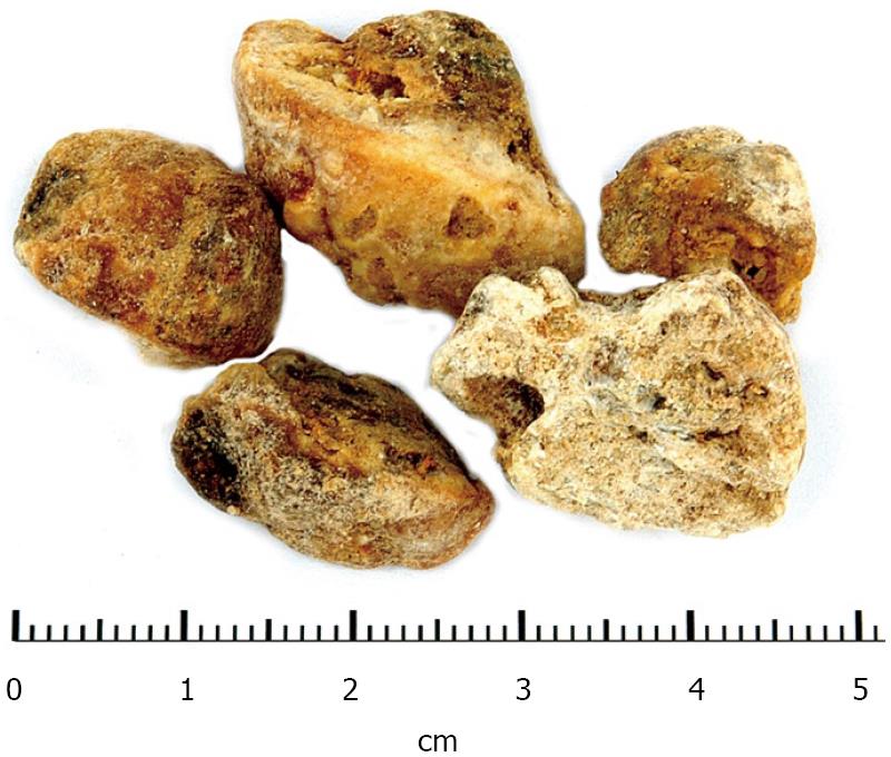
Because of the poor physical condition and underlying comorbidities of the patient, surgery was considered to carry too high a risk. Therefore, single-balloon enteroscopy (SBE, SIF-100 enteroscope, Olympus, Japan) via the oral route was performed revealing a calculus about 450 cm distant from the pylorus, completely occluding the intestinal lumen (Figure 3). Under endoscopic guidance the calculus was captured with a Dormia basket but could not be retracted due to intestinal incarceration. Therefore the endoscope was withdrawn leaving the captured calculus in situ and leading out the basket wire pernasally. In order to facilitate calculus removal three sessions of ESWL (4000 pulses each session) were performed. Thereafter, the single-balloon enteroscope was reinserted via the oral route guided by the wire of the Dormia basket. This time, under fluoroscopic control (Figure 4) the partially disintegrated calculus could be endoscopically retrieved up to the stomach. Since the size of the calculus did not allow retraction through the lower esophageal sphincter, endoscopically guided laser lithotripsy (Calculase 27750120 Desktop Holmium YAG Laser, Karl Storz, Germany) and mechanical fragmentation were performed permitting safe peroral endoscopic removal of all gallstone fragments (Figure 5). 3 d later the patient was discharged in good health. Endoscopic follow-up at 4 wk after discharge revealed the deep duodenal bulb ulcer to be healed. Neither pneumobilia nor any fistulous structure could be observed radiographically.
Gallstone-induced ileus is a rare complication of cholelithiasis and is associated with relatively high rates of mortality[2]. Endoscopic removal of gallstones from the upper jejunum has been described[9]. However, since localization of gallstones impacted in the small bowel prevents access of conventional endoscopy in most cases, such situations usually require surgical treatment[1-3]. Recent invention of double- and single-balloon enteroscopy[10,11] has added much to the ability of imaging the small bowel and enables endoscopically directed therapy such as polypectomy, extraction of foreign bodies and arrest of bleeding. In a study by Ramchandani and colleagues with 106 patients the mean insertion depth via SBE was 255.8 cm ± 84.5 cm beyond the duodenojejunal flexure by the oral route and 163 cm ± 59.3 cm proximal to the ileocecal valve by the peranal approach[12]. Pan-enteroscopy is possible in 25% to 60% of cases combining peroral and peranal access[11,12] and is a highly useful diagnostic and therapeutic tool in endoscopy[12]. Therefore, the use of SBE to detect jejunal or ileal gallstones appears promising.
Consequently, in the present setting, single-balloon enteroscopy was performed and proved to be a successful minimally-invasive and safe method for calculus removal.
This report for the first time demonstrates the beneficial role of single-balloon enteroscopy in treating small bowel gallstone ileus, potentially obviating the necessity of surgical intervention. Therefore, in cases similar to that reported here minimal invasive SBE should be the primary therapeutic approach with the surgical option still remaining in case of failure. The development of special probes for laser lithotripsy via SBE would be ideal. As long as such probes are not available, however, we recommend the combined procedure of SBE and ESWL.
Peer reviewers: Sherman M Chamberlain, MD, FACP, FACG, AGAF, Associate Professor of Medicine, Section of Gastroenterology, BBR-2538, Medical College of Georgia, Augusta, GA 30912, United States; Iruru Maetani, MD, Professor and Chairman, Division of Gastroenterology, Department of Internal Medicine, Toho University Ohashi Medical Center, 2-17-6 Ohashi Meguro-ku, Tokyo 153-8515, Japan
S- Editor Zhang HN L- Editor Hughes D E- Editor Liu N
| 1. | Day EA, Marks C. Gallstone ileus. Review of the literature and presentation of thirty-four new cases. Am J Surg. 1975;129:552-558. |
| 2. | Reisner RM, Cohen JR. Gallstone ileus: a review of 1001 reported cases. Am Surg. 1994;60:441-446. |
| 3. | van Hillo M, van der Vliet JA, Wiggers T, Obertop H, Terpstra OT, Greep JM. Gallstone obstruction of the intestine: an analysis of ten patients and a review of the literature. Surgery. 1987;101:273-276. |
| 4. | Deitz DM, Standage BA, Pinson CW, McConnell DB, Krippaehne WW. Improving the outcome in gallstone ileus. Am J Surg. 1986;151:572-576. |
| 5. | Sackmann M, Holl J, Haerlin M, Sauerbruch T, Hoermann R, Heinkelein J, Paumgartner G. Gallstone ileus successfully treated by shock-wave lithotripsy. Dig Dis Sci. 1991;36:1794-1795. |
| 6. | Fujita N, Noda Y, Kobayashi G, Kimura K, Watanabe H, Shirane A, Hayasaka T, Mochizuki F, Yamazaki T. Gallstone ileus treated by electrohydraulic lithotripsy. Gastrointest Endosc. 1992;38:617-619. |
| 7. | Zielinski MD, Ferreira LE, Baron TH. Successful endoscopic treatment of colonic gallstone ileus using electrohydraulic lithotripsy. World J Gastroenterol. 2010;16:1533-1536. |
| 8. | Meyenberger C, Michel C, Metzger U, Koelz HR. Gallstone ileus treated by extracorporeal shockwave lithotripsy. Gastrointest Endosc. 1996;43:508-511. |
| 9. | Lubbers H, Mahlke R and Lankisch PG. Gallstone ileus: endoscopic removal of a gallstone obstructing the upper jejunum. J Intern Med. 1999;246:593-597. |
| 10. | Yamamoto H, Sekine Y, Sato Y, Higashizawa T, Miyata T, Iino S, Ido K, Sugano K. Total enteroscopy with a nonsurgical steerable double-balloon method. Gastrointest Endosc. 2001;53:216-220. |
| 11. | Kobayashi K, Haruki S, Sada M, Katsumata T, Saigenji K. [Single-balloon enteroscopy].. Nippon Rinsho. 2008;66:1371-1378. |
| 12. | Ramchandani M, Reddy DN, Gupta R, Lakhtakia S, Tandan M, Rao GV, Darisetty S. Diagnostic yield and therapeutic impact of single-balloon enteroscopy: series of 106 cases. J Gastroenterol Hepatol. 2009;24:1631-1638. |


