Published online Jul 16, 2010. doi: 10.4253/wjge.v2.i7.268
Revised: June 26, 2010
Accepted: July 3, 2010
Published online: July 16, 2010
Colocolonic intussusception is an uncommon cause of intestinal obstruction in children. The most common type is idiopathic ileocolic intussusception. However, pathologic lead points occur approximately in 5% of cases. In pediatric patients, Meckel’s diverticulum is the most common lead point, followed by polyps and duplication. We present a case of recurrent colocolonic intussusception which caused colonic obstruction in a 10-year-old boy. A barium enema revealed a large polypoid mass at the transverse colon. Colonoscopy showed a colonic polyp, 3.5 centimeters in diameter, which was successfully removed by endoscopic polypectomy.
- Citation: Suksamanapun N, Uiprasertkul M, Ruangtrakool R, Akaraviputh T. Endoscopic treatment of a large colonic polyp as a cause of colocolonic intussusception in a child. World J Gastrointest Endosc 2010; 2(7): 268-270
- URL: https://www.wjgnet.com/1948-5190/full/v2/i7/268.htm
- DOI: https://dx.doi.org/10.4253/wjge.v2.i7.268
Colocolonic intussusception is an uncommon cause of intestinal obstruction in children[1,2]. The most common type is idiopathic ileocolic intussusception. On rare occasions, in less than 3% of cases, colocolonic intussusception occurs and is usually associated with no pathologic lead point[3,4].
The pathologic lead point occurs in only 5% of intussusception cases, and is usually present in older children[1,5]. The most common pathologic lead point is Meckel’s diverticulum, followed by small bowel polyps and intestinal duplications[2,6].
We present a case of colocolonic intussusception in a 10-year-old boy. The patient had as pathologic lead point a large polyp which was completely removed by endoscopic surgery. The pathological result confirmed a juvenile colonic polyp.
A 10-year-old boy was admitted to the Division of Pediatric Surgery, Department of Surgery, Faculty of Medicine at Siriraj Hospital due to recurrent episodes of intussusception. In a provincial hospital, he presented with a one-day history of colicky abdominal pain and bloody stool. Abdominal ultrasonography was performed, and confirmed the diagnosis of intussusception. After hydrostatic reduction failed, he underwent a laparotomy for manual reduction. The patient recovered and was discharged from the hospital on the day 5.
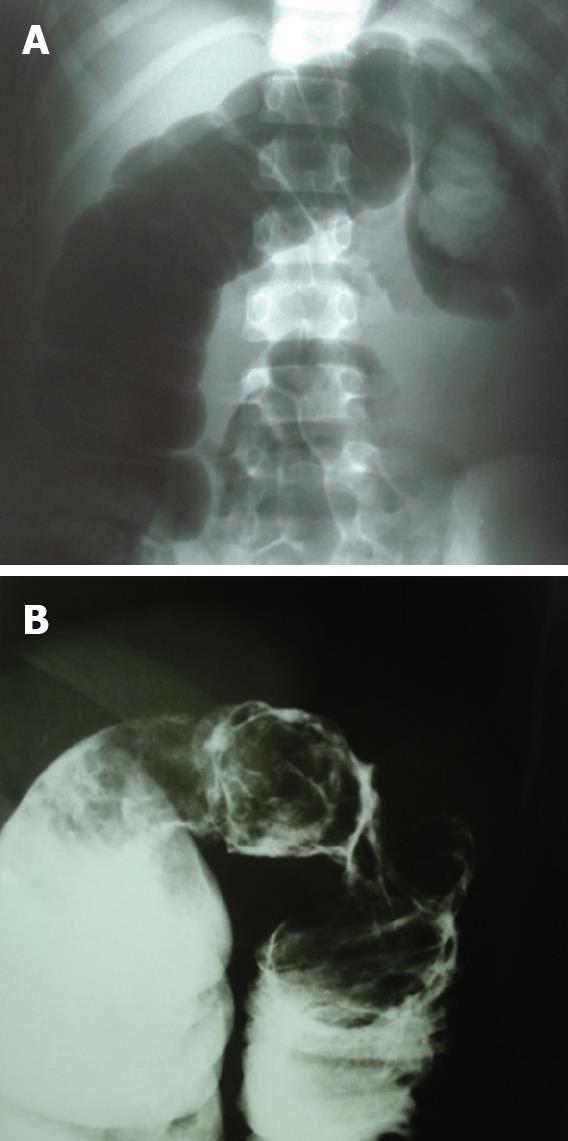
8 d after the operation, colicky pain recurred as well as the bloody stool. A plain abdominal film showed colonic obstruction near the splenic flexure of the colon (Figure 1A). Colocolonic intussusception was successfully reduced by hydrostatic pressure. A barium enema revealed a mucosal lesion at the splenic flexure of the colon (Figure 1B). The patient was then referred to our hospital.
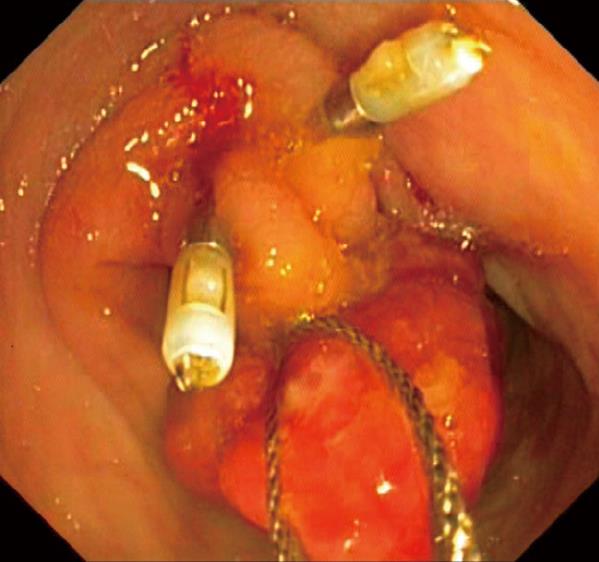
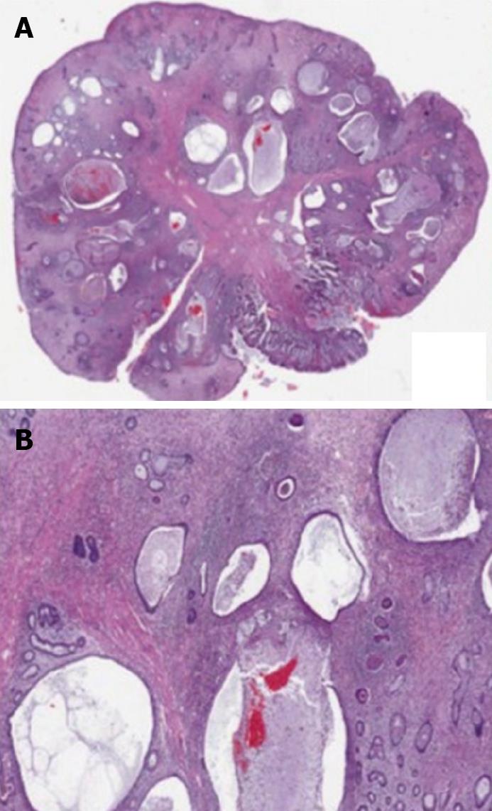
He underwent an elective colonoscopy 1 wk later. A large pedunculated polyp, measuring 3.5 centimeters in diameter, was detected at the transverse colon and polypectomy was successfully performed (Figure 2). The pathologic finding revealed a juvenile polyp (Figure 3). The child recovered and has done well ever since.
Although intussusception has been reported in all pediatric age groups, 75%-90% of the cases occur within the first 2 years of life[6]. Most of the presentations of intussusception are of the idiopathic ileocolic type[4,6,7]. Only 5%-6% of these patients have pathologic lead points[1,2,4]. The incidence of pathologic lead points increases with age and recurrent episodes. Intussusception which occurs outside the ileocolic junction appears to have a higher incidence of pathologic lead points and requires surgical management such as a bowel resection[4]. This patient should have been initially suspected to have a pathological lead point with the development of recurrent colocolonic intussusception.
The approach to patients with intussusception due to a pathological lead point is similar to that of patients in the idiopathic group, as it may at first be difficult to diagnose them from standard management procedures, such as presenting history, physical examination and a plain film. A contrast enema reduction is the first line of the management, unless the patient shows signs of secondary peritonitis. Lead points which are caused by diffuse mucosal lesions benefit from this approach. Some of them (non-pathological lead points) are associated with inherent existing conditions such as hereditary angioneurotic edema[8]. A careful review of post-evacuation films should be performed in cases where there is a strong possibility of a pathologic lead point[9]. In the colocolonic intussusceptions group, colonoscopy is indicated in order to identify a lead point in the colon. Endoscopic treatment, such as polypectomy, can safely be performed when removable mucosal lesions are present[3,10]. This intervention can reduce unnecessary or invasive surgery. However, pediatric colonoscopy requires sedation and/or a general anesthetic, as opposed to a barium enema which, in good pediatric radiology hands, can be just as effective for diagnosis and further information.
A juvenile colonic polyp can act as a lead point for colocolonic intussusception[3]. Juvenile polyps usually occur in first decade of life. The usual presenting symptom is painless hematochezia[11,12]. Although a solitary lesion is often benign, malignant transformation can occur in juvenile polyposis syndrome when the number of juvenile polyps is greater than 5[12]. A complete colonoscopy plays a crucial role in distinguishing juvenile polyposis syndrome from a solitary polyp. Surveillance colonoscopy is recommended in those who have multiple polyps and precancerous lesions.
In conclusion, recurrent colocolonic intussusception in a school-age patient is rare and usually due to a pathologic lead point. Goals for the treatment are the reduction of intussusception and searching for and finding a lead point. Radiologic reduction is a useful initial treatment. However, it is seldom successful in reducing a colocolic intussusception, an intussusception in an older child, and/or a recurrent intussusception, all of which are symptoms of an intussusception caused by a pathological lead point. A careful review of post-evacuation films is helpful. Colonoscopy is a non-invasive and effective tool in searching for intraluminal lesions in the colon such as polyps, which can be simultaneously removed.
Peer reviewer: Omar Javed Shah, Professor, Head, Department of Surgical Gastroenterology, Sher-i-Kashmir Institute of Medical Sciences, Srinagar, Kashmir 190011, India
| 1. | Applegate KE. Intussusception in children: evidence-based diagnosis and treatment. Pediatr Radiol. 2009;39 Suppl 2:S140-S143. |
| 2. | Chung JL, Kong MS, Lin JN, Wang KL, Lou CC, Wong HF. Intussusception in infants and children: risk factors leading to surgical reduction. J Formos Med Assoc. 1994;93:481-485. |
| 3. | Arthur AL, Garvey R, Vaness DG. Colocolic intussusception in a three-year-old child caused by a colonic polyp. Conn Med. 1990;54:492-494. |
| 4. | Chua JH, Chui CH, Jacobsen AS. Role of surgery in the era of highly successful air enema reduction of intussusception. Asian J Surg. 2006;29:267-273. |
| 5. | Soccorso G, Puls F, Richards C, Pringle H, Nour S. A ganglioneuroma of the sigmoid colon presenting as leading point of intussusception in a child: a case report. J Pediatr Surg. 2009;44:e17-e20. |
| 6. | Waseem M, Rosenberg HK. Intussusception. Pediatr Emerg Care. 2008;24:793-800. |
| 7. | Grant HW, Buccimazza I, Hadley GP. A comparison of colo-colic and ileo-colic intussusception. J Pediatr Surg. 1996;31:1607-1610. |
| 8. | Pritzker HA, Levin TL, Weinberg G. Recurrent colocolic intussusception in a child with hereditary angioneurotic edema: reduction by air enema. J Pediatr Surg. 2004;39:1144-1146. |
| 9. | Ong NT, Beasley SW. The leadpoint in intussusception. J Pediatr Surg. 1990;25:640-643. |
| 10. | Caputi IO, Ugenti I, Martines G, Marino F, Francesco AD, Memeo V. Endoscopic management of large colorectal polyps. Int J Colorectal Dis. 2009;24:749-753. |
| 11. | Ukarapol N, Singhavejakul J, Lertprasertsuk N, Wongsawasdi L. Juvenile polyp in Thai children--clinical and colonoscopic presentation. World J Surg. 2007;31:395-398. |
| 12. | Haghi AMT, Monajemzadeh M, Motamed F, Moradi TH, Mahjoub F, Karamian H, Najafi SM, Khatami GR, Khodadad A, Farahmand F. Colorectal polyps: a clinical, endoscopic and pathologic study in Iranian children. Med Princ Pract. 2009;18:53-56. |


