Published online Nov 16, 2020. doi: 10.4253/wjge.v12.i11.451
Peer-review started: July 16, 2020
First decision: September 14, 2020
Revised: September 29, 2020
Accepted: October 20, 2020
Article in press: October 20, 2020
Published online: November 16, 2020
Processing time: 122 Days and 23.9 Hours
Compared to traditional open surgery, laparoscopic surgery has become a standard approach for colorectal cancer due to its great superiorities including less postoperative pain, a shorter hospital stay, and better quality of life. In 2007, Whiteford et al reported the first natural orifice trans-anal endoscopic surgery (NOTES) sigmoidectomy using transanal endoscopic microsurgery. To date, all cases of NOTES colorectal resection have included a hybrid laparoscopic approach with the use of established rigid platforms.
To introduce a novel technique of peroral external traction-assisted transanal NOTES rectosigmoidectomy followed by intracorporeal colorectal end-to-end anastomosis by using only currently available and flexible endoscopic instrumentation in a live porcine model.
Three female pigs weighing 25-30 kg underwent NOTES rectosigmoid resection. After preoperative work-up and bowel preparation, general anesthesia combined with endotracheal intubation was achieved. One dual-channel therapeutic endoscope was used. Carbon dioxide insufflation was performed during the operation. The procedure of trans-anal NOTES rectosigmoidectomy included the following eight steps: (1) The rectosigmoid colon was tattooed with India ink by submucosal injection; (2) Creation of gastrostomy by directed submucosal tunneling; (3) Peroral external traction using endoloop ligation; (4) Creation of rectostomy on the anterior rectal wall by directed 3 cm submucosal tunneling; (5) Peroral external traction-assisted dissection of the left side of the colon; (6) Trans-anal rectosigmoid specimen transection, where an anvil was inserted into the proximal segment after purse-string suturing; (7) Intracorporeal colorectal end-to-end anastomosis using a circular stapler by a single stapling technique; and (8) Closure of gastrostomy using endoscopic clips. All animals were euthanized immediately after the procedure, abdominal exploration was performed, and the air-under-water leak test was carried out.
The procedure was completed in all three animals, with the operation time ranging from 193 min to 259 min. Neither major intraoperative complications nor hemodynamic instability occurred during the operation. The length of the resected specimen ranged from 7 cm to 13 cm. With the assistance of a trans-umbilical rigid grasper, intracorporeal colorectal, tension-free, end-to-end anastomosis was achieved in the three animals.
Peroral traction-assisted transanal NOTES rectosigmoidectomy followed by intracorporeal colorectal end-to-end anastomosis is technically feasible and reproducible in an animal model and is worthy of further improvements.
Core Tip: A novel technique, natural orifice trans-anal endoscopic (NOTES) rectosigmoidectomy followed by intracorporeal colorectal end-to-end anastomosis, may be successfully performed in a live porcine model with the assistance of peroral external traction and the trans-umbilical rigid grasper.
- Citation: Shi H, Chen SY, Xie ZF, Huang R, Jiang JL, Lin J, Dong FF, Xu JX, Fang ZL, Bai JJ, Luo B. Peroral traction-assisted natural orifice trans-anal flexible endoscopic rectosigmoidectomy followed by intracorporeal colorectal anastomosis in a live porcine model. World J Gastrointest Endosc 2020; 12(11): 451-458
- URL: https://www.wjgnet.com/1948-5190/full/v12/i11/451.htm
- DOI: https://dx.doi.org/10.4253/wjge.v12.i11.451
Compared to traditional open surgery, laparoscopic surgery has become a standard approach for colorectal cancer due to its great superiorities including less postoperative pain, a shorter hospital stay, and better quality of life. Since the first report of its clinical application in 1991, an increasing number of minimally invasive surgical techniques, including single-incision laparoscopic surgery (SILS)[1], needlescopic surgery (NS)[2], and natural orifice transluminal endoscopic surgery (NOTES)[3], have been developed rapidly. Of these, only NOTES can provide an opportunity for incision-free abdominal surgery. Although NOTES-related techniques continue to evolve, they remain mainly confined to animal models due to technical constraints and instrument limitations. In 2007, Whiteford et al[4] reported the first NOTES sigmoidectomy using transanal endoscopic microsurgery. To date, all cases of NOTES colorectal resection have included a hybrid laparoscopic approach with the use of established rigid platforms. Our study aimed to introduce the novel technique of peroral external traction-assisted transanal NOTES sigmoidectomy followed by intracorporeal colorectal end-to-end anastomosis by using only currently available and flexible endoscopic instrumentation in a live swine model.
Three female pigs weighing 25-30 kg were used in this study. Preoperative work-up and bowel preparation comprised a 3-d liquid diet and a 1-d fast, followed by preoperative polyethylene glycol given orally. The induction of anesthesia was achieved by an intramuscular injection of 100 mg ketamine, 10 mg droperidol, and 1 mg atropine, and the maintenance of anesthesia was achieved by an intravenous drip of propofol at a dosage of 10 mL/h after endotracheal intubation. The heart rate and oxygen saturation of each animal were monitored during the operation. Animals were maintained in a supine Trendelenburg position to allow for optimal access and peritoneal exploration[5]. One dual-channel therapeutic endoscope (GIF-2TQ260M, Olympus) was used. Carbon dioxide insufflation was performed during the operation. This study was approved by the Institutional Animal Use and Care Committee of Beijing Pinggu Hospital and Fuzhou General Hospital of Nanjing Military Command (IACUC-2015-010).
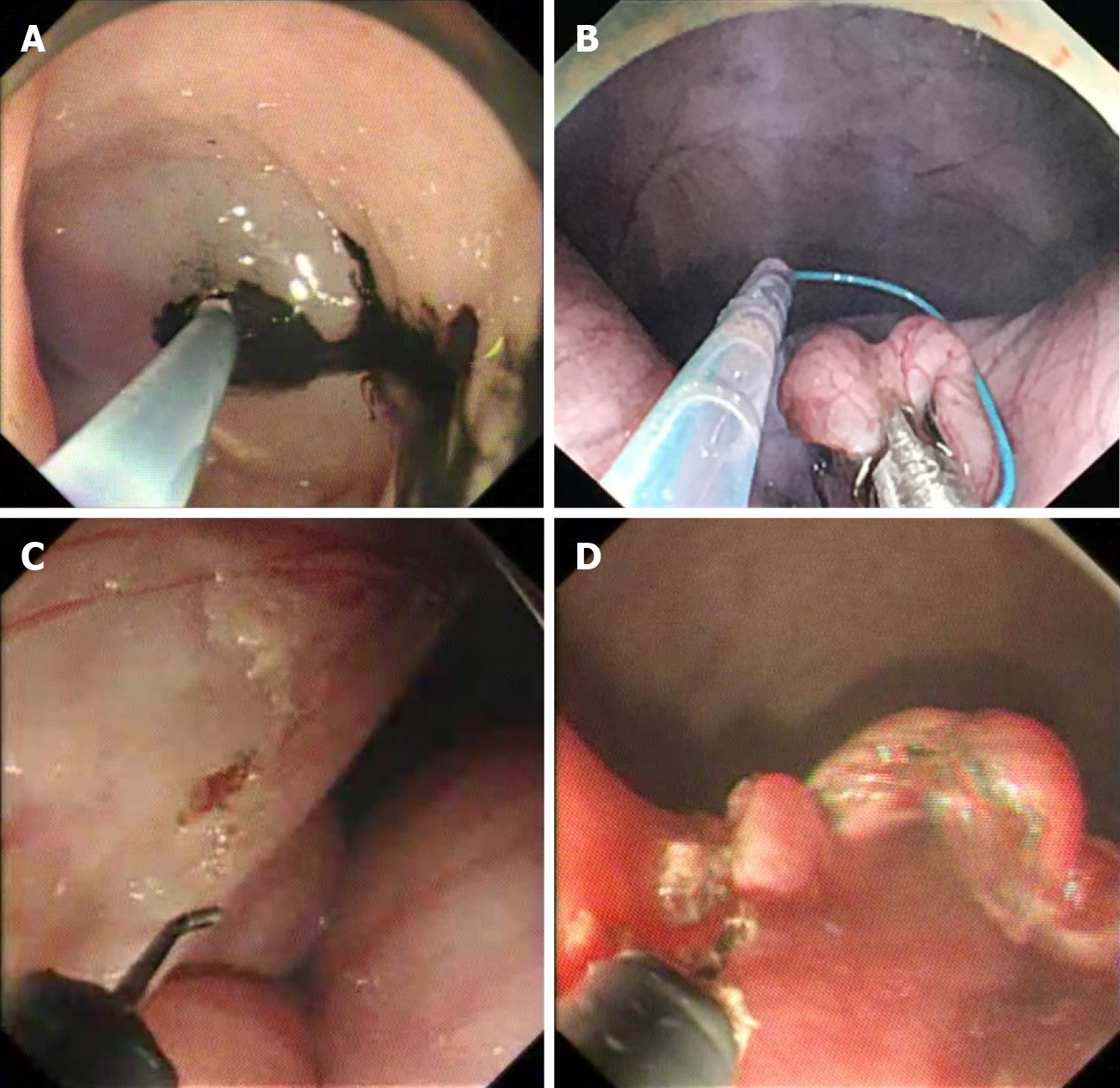
The anterior wall of the rectosigmoid colon was tattooed with India ink by submucosal injection under trans-anal endoscopic vision (Figures 1 and 2).
Creation of gastrostomy by directed submucosal tunneling under trans-gastric endoscopic vision[5]: A 2-cm transversal mucosal incision was created near the gastroesophageal junction with a dual knife (KD650L; Olympus, Tokyo Japan), followed by the creation of a 3-5 cm longitudinal submucosal pelvis-directed tunnel. The tunnel ended with a seromuscular incision, and the exit site was selected at the anterior wall of the stomach.
Peroral external traction using endoloop ligation under trans-gastric endoscopic vision: An external endoloop knotted to a segment of dental floss was passed through by a twin grasper[6] via one of the accessory channels of the endoscope. Then, the dual-channel therapeutic endoscope was again advanced into the peritoneal cavity through the gastrostomy site. After abdominal exploration, the twin grasper was used to catch and pull the anterior wall of the rectosigmoid colon tattooed with India ink so that the endoloop might rope the part of the targeted colon. Once the endoloop was tightened followed by stretching of the floss, peroral external traction was achieved, allowing exposure of the sigmoid mesocolon and subsequent endoscopic dissection of the vessel and mesentery.
Creation of rectostomy on the anterior rectal wall 5 cm distal to the tattooed marker of the rectosigmoid colon by using directed short submucosal tunneling under trans-anal endoscopic vision: This was the same as the creation of gastrostomy.
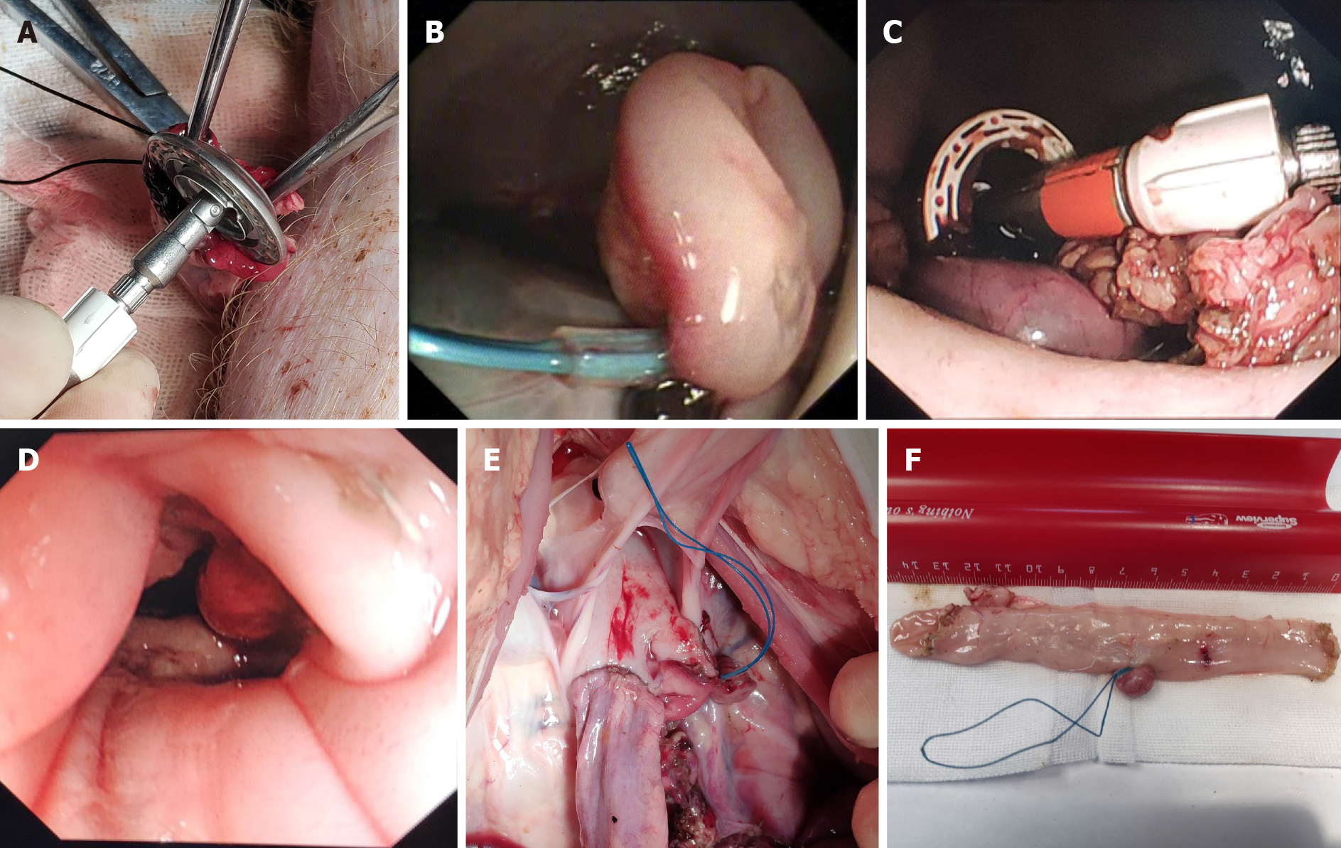
Peroral external traction-assisted dissection of the left side of the colon under trans-anal endoscopic vision: With the help of peroral external traction, the sigmoid colon mesentery was mobilized off the retroperitoneum with a hook knife (model KD-620LR; Olympus). The inferior mesenteric vessels were successfully dissected using a Coagrasper (model FD-410LR; Olympus) and endoscopic clips (HX-600-135; Olympus), which was similar to the description issued by Park et al[7]. After being dissected for around ten cm in length, the mobilized rectosigmoid colonic segment was transected at the site of the tunnel entrance.
Trans-anal rectosigmoid specimen transection: The mobilized rectosigmoid colon was exteriorized and transected trans-anally. A 25-mm circular stapler anvil (Medtronic) was inserted into the proximal segment after purse-string suturing, and the proximal bowel was then returned into the abdomen.
Intracorporeal end-to-end colorectal anastomosis using a circular stapler by a single stapling technique under trans-gastric endoscopic guidance: The dual-channel therapeutic endoscope was again advanced into the peritoneal cavity through the gastrostomy site. Pneumoperitoneum was reestablished, and then an endoloop was used to ligate the lateral rectostomy by endoscopy. After the stapler was inserted into the rectum and pricked the top wall of the rectum, a trans-umbilical rigid grasper was used to orient the proximal bowel properly and then guide the proximal stapler anvil to mate with the stapler. Once apposed, the stapler was fired. The stapler was then removed, and the anastomotic tissue rings were immediately inspected for completeness by trans-anal endoscopy.
Closure of gastrostomy using endoscopic clips under trans-gastric endoscopic vision: The defect of the gastric tunnel entrance was closed with endoscopic clips.
After the procedure, all three animals were euthanized immediately, abdominal exploration was performed, and the air-under-water leak test was carried out[4]. The pelvis was filled with normal saline, and the rectum was insufflated to confirm whether the anastomosis was airtight.
The primary outcome of this study was the procedure success rate. The secondary outcomes were the total operative time, specimen length, completeness of colorectal anastomosis, and adverse event rate in the perioperative period. At necropsy, the anastomosis was tested for leaks using the air-under-water test.
The animal protocol was designed to minimize pain or discomfort to the animals. All animals were euthanized by barbiturate overdose (intravenous injection, 150 mg/kg pentobarbital sodium) for autopsy.
The procedure was completed in all three animals, with the total operation time ranging from 193 min to 259 min (Table 1). Neither intraoperative complications nor hemodynamic instability occurred during the operation. Adequate anatomic exposure around the inferior mesenteric vessels was achieved by peroral external traction using endoloop ligation. Endoscopic dissection of the inferior mesenteric vessels was successful in all cases. The length of the resected specimen ranged from 7 cm to 13 cm, attached by the sigmoid mesentery.
| Animal No. | Rectosigmoidectomy | Specimen length (cm) | Duration (min) | Anastomosis completeness |
| 1 | Success | 9 | 259 | Complete |
| 2 | Success | 13 | 217 | Uncomplete |
| 3 | Success | 7 | 193 | Complete |
With the assistance of a trans-umbilical rigid grasper, intracorporeal end-to-end colorectal anastomosis was achieved in all three animals. The anastomotic tissue ring in the second case was noted to be incomplete along the posterior rectal wall due to the insufficient occluding purse-string suturing of the proximal colonic segment. This may be a result of excessive resection of the sigmoid colon leading to retraction of the proximal segment, impairing sufficient purse-string suturing. The anastomotic defect was then reinforced with clips by tans-anal endoscopy.
At necropsy, there were no injuries to the adjacent organs. A properly oriented, tension-free colorectal end-to-end anastomosis was achieved in all three animals. Fortunately, the leak test was also negative in all animals regardless of whether anastomotic completeness was achieved.
To the best of our knowledge, this is the first study assessing the feasibility and safety of peroral external traction-assisted transanal NOTES sigmoidectomy followed by intracorporeal colorectal end-to-end anastomosis by using only currently available endoscopic flexible accessories except a rigid grasper in a live swine model.
In our study, endoscopic sigmoid mesocolon dissection, major vessel ligation, and en bloc retrieval were feasible via the pure NOTES approach. As Park et al[7] stated, “The most important is that the operating field exposure through traction should be performed before dissection itself”. Different from the description of Park et al[7], in our study, external traction of the sigmoid mesocolon was achieved through trans-oral introduction of an endoloop knotted to a segment of dental floss. Furthermore, our traction method could be used in the whole colon, while traction through the trans-anal introduction of a circular stapler was only available for the sigmoid colon[7]. Notably, the direction of traction was fixed both in our study and Park SJ’s study. In the future, gastrointestinal endoscopic robots may enable real-time changes in traction direction by remote control[8].
The CO2 pneumoperitoneum maintained by an endoscopic insufflator also permitted intra-abdominal visualization. Since it was difficult to monitor the intra-abdominal pressure during the procedure, endoscopic discontinuous suction was necessary.
In contrast to extracorporeal colorectal anastomosis published in previous reports[9-13], intracorporeal end-to-end anastomosis under trans-gastric endoscopic guidance was introduced in our study to achieve high colorectal anastomosis. According to the updated metaanalysis, compared to extracorporeal anastomosis, intracorporeal anastomosis may be associated with a shorter extraction site incision, faster bowel recovery, fewer perioperative complications, and lower rates of conversion to open surgery, anastomotic leakage, surgical site infection, and incisional hernia[14-18]. In our study, the most technically challenging and time-consuming step was to mate the proximal stapler anvil with the stapler inserted trans-anally. A trans-umbilical rigid grasper was used to achieve alignment.
Similar to gastrostomy, lateral rectostomy on the anterior rectal wall was achieved by using directed short submucosal tunneling for subsequent end-to-end anastomotic creation.
To date, gastric closure remains one of the major difficulties, and endoscopic clipping can only achieve mucosal apposition. For secure gastric closure, the creation of gastrostomy by directed submucosal tunneling was applied in this study so that we only needed to close the mucosal defect of the gastric tunnel entrance[19].
There were also several technical challenges in our study. First, due to the lack of a wide field of vision and the spatial orientation of laparoscopy, accurate endoscopic dissection is still technically demanding. It is difficult to precisely identify the beginning and endpoint of the colon segment to be dissected. Since virtual reality with three-dimensional reconstruction allows an enhanced understanding of crucial anatomical details, it would contribute to improving safety and accuracy in endoscopic surgery[20-23]. Second, although intracorporeal end-to-end anastomosis was achieved in this study, rigid instrumentation was still needed. Before clinical application of this technique, instrument development, including endoscopic anastomotic equipment, would be required[24-26]. Third, to determine whether the anastomotic method can achieve histological anastomosis, subsequent survival experiments should be carried out.
In conclusion, this novel technique for performing NOTES sigmoidectomy with the assistance of peroral external traction, followed by intracorporeal colorectal end-to-end anastomosis aided by a trans-umbilical rigid grasper, is safe and feasible in a live animal model and is worthy of further improvements.
Since 1991, an increasing number of minimally invasive surgical techniques, including single-incision laparoscopic surgery (SILS), needlescopic surgery (NS), and natural orifice transluminal endoscopic surgery (NOTES), have been developed rapidly. To date, all cases of NOTES colorectal resection have included a hybrid laparoscopic approach with the use of established rigid platforms.
Our research aimed to improve NOTES-related techniques.
Our study aimed to introduce the novel technique of peroral external traction-assisted transanal NOTES sigmoidectomy followed by intracorporeal colorectal end-to-end anastomosis by using only currently available and flexible endoscopic instrumentation in a live swine model.
Three female pigs weighing 25-30 kg underwent NOTES rectosigmoid resection. The procedure of trans-anal NOTES rectosigmoidectomy included the following eight steps: (1) The rectosigmoid colon was tattooed with India ink by submucosal injection; (2) Creation of gastrostomy by directed submucosal tunneling; (3) Peroral external traction using endoloop ligation; (4) Creation of rectostomy on the anterior rectal wall by directed 3 cm submucosal tunneling; (5) Peroral external traction-assisted dissection of the left side of the colon; (6) Trans-anal rectosigmoid specimen transection, where an anvil was inserted into the proximal segment after purse-string suturing; (7) Intracorporeal colorectal end-to-end anastomosis using a circular stapler with a single stapling technique; and (8) Closure of gastrostomy using endoscopic clips.
The procedure was completed in all three animals, with the operation time ranging from 193 min to 259 min. The length of the resected specimen ranged from 7 cm to 13 cm. With the assistance of a trans-umbilical rigid grasper, intracorporeal colorectal, tension-free, end-to-end anastomosis was achieved in the three animals.
Peroral traction-assisted transanal NOTES rectosigmoidectomy followed by intracorporeal colorectal end-to-end anastomosis is technically feasible and reproducible in an animal model and is worthy of further improvements.
The techniques of NOTES rectosigmoidectomy need to be improved for clinical application.
Manuscript source: Unsolicited manuscript
Corresponding Author's Membership in Professional Societies: The Vice Chairman of Committee of Society of Tumor Endoscopy, Chinese Anti-Cancer Association; The Chairman of Committee of Society of Tumor Endoscopy, Fujian Anti-Cancer Association; The Director of ESD Training Center, Chinese Medical Doctor Association; The Standing Committee of Society of Fujian Gastrointestinal Endoscopy; and Member of Europe Society of Gastrointestinal Endoscopy.
Specialty type: Gastroenterology and hepatology
Country/Territory of origin: China
Peer-review report’s scientific quality classification
Grade A (Excellent): 0
Grade B (Very good): B
Grade C (Good): C
Grade D (Fair): 0
Grade E (Poor): 0
P-Reviewer: Shichijo S, Sozutek A S-Editor: Gao CC L-Editor: Wang TQ P-Editor: Liu JH
| 1. | Hirano Y, Hiranuma C, Hattori M, Douden K, Yamaguchi S. Single-incision or Single-incision Plus One-Port Laparoscopic Surgery for Colorectal Cancer. Surg Technol Int. 2020;36:132-135. [PubMed] |
| 2. | Miki H, Fukunaga Y, Nagasaki T, Akiyoshi T, Konishi T, Fujimoto Y, Nagayama S, Ueno M. Feasibility of needlescopic surgery for colorectal cancer: safety and learning curve for Japanese Endoscopic Surgical Skill Qualification System-unqualified young surgeons. Surg Endosc. 2020;34:752-757. [RCA] [PubMed] [DOI] [Full Text] [Cited by in Crossref: 4] [Cited by in RCA: 9] [Article Influence: 1.5] [Reference Citation Analysis (0)] |
| 3. | Buscaglia JM, Karas J, Palladino N, Fakhoury J, Denoya PI, Nagula S, Bucobo JC, Bishawi M, Bergamaschi R. Simulated transanal NOTES sigmoidectomy training improves the responsiveness of surgical endoscopists. Gastrointest Endosc. 2014;80:126-132. [RCA] [PubMed] [DOI] [Full Text] [Cited by in Crossref: 4] [Cited by in RCA: 6] [Article Influence: 0.5] [Reference Citation Analysis (0)] |
| 4. | Whiteford MH, Denk PM, Swanström LL. Feasibility of radical sigmoid colectomy performed as natural orifice translumenal endoscopic surgery (NOTES) using transanal endoscopic microsurgery. Surg Endosc. 2007;21:1870-1874. [RCA] [PubMed] [DOI] [Full Text] [Cited by in Crossref: 222] [Cited by in RCA: 198] [Article Influence: 11.0] [Reference Citation Analysis (0)] |
| 5. | Shi H, Chen SY, Wang YG, Jiang SJ, Cai HL, Lin K, Xie ZF, Dong FF. Percutaneous transgastric endoscopic tube ileostomy in a porcine survival model. World J Gastroenterol. 2016;22:8375-8381. [RCA] [PubMed] [DOI] [Full Text] [Full Text (PDF)] [Cited by in CrossRef: 1] [Cited by in RCA: 1] [Article Influence: 0.1] [Reference Citation Analysis (1)] |
| 6. | Kobara H, Mori H, Fujihara S, Nishiyama N, Chiyo T, Yamada T, Fujiwara M, Okano K, Suzuki Y, Murota M, Ikeda Y, Oryu M, AboEllail M, Masaki T. Outcomes of gastrointestinal defect closure with an over-the-scope clip system in a multicenter experience: An analysis of a successful suction method. World J Gastroenterol. 2017;23:1645-1656. [RCA] [PubMed] [DOI] [Full Text] [Full Text (PDF)] [Cited by in CrossRef: 30] [Cited by in RCA: 30] [Article Influence: 3.8] [Reference Citation Analysis (0)] |
| 7. | Park SJ, Lee KY, Choi SI, Kang BM, Huh C, Choi DH, Lee CK. Pure NOTES rectosigmoid resection: transgastric endoscopic IMA dissection and transanal rectal mobilization in animal models. J Laparoendosc Adv Surg Tech A. 2013;23:592-595. [RCA] [PubMed] [DOI] [Full Text] [Cited by in Crossref: 3] [Cited by in RCA: 3] [Article Influence: 0.3] [Reference Citation Analysis (0)] |
| 8. | Kume K, Sakai N, Ueda T. Development of a Novel Gastrointestinal Endoscopic Robot Enabling Complete Remote Control of All Operations: Endoscopic Therapeutic Robot System (ETRS). Gastroenterol Res Pract. 2019;2019:6909547. [RCA] [PubMed] [DOI] [Full Text] [Full Text (PDF)] [Cited by in Crossref: 10] [Cited by in RCA: 10] [Article Influence: 1.7] [Reference Citation Analysis (0)] |
| 9. | Wilhelm P, Axt S, Storz P, Wenz S, Müller S, Kirschniak A. Pure Natural Orifice Transluminal Endoscopic Surgery (NOTES) with a new elongated, curved Transanal Endoscopic Operation (TEO) device for rectosigmoid resection: a survival study in a porcine model. Tech Coloproctol. 2016;20:273-278. [RCA] [PubMed] [DOI] [Full Text] [Cited by in Crossref: 4] [Cited by in RCA: 4] [Article Influence: 0.4] [Reference Citation Analysis (0)] |
| 10. | Park SJ, Sohn DK, Chang TY, Jung Y, Kim HJ, Kim YI, Chun HK; Korea Natural Orifice Transluminal Endoscopic Surgery (K-NOTES) Study Group. Transanal natural orifice transluminal endoscopic surgery total mesorectal excision in animal models: endoscopic inferior mesenteric artery dissection made easier by a retroperitoneal approach. Ann Surg Treat Res. 2014;87:1-4. [RCA] [PubMed] [DOI] [Full Text] [Full Text (PDF)] [Cited by in Crossref: 5] [Cited by in RCA: 3] [Article Influence: 0.3] [Reference Citation Analysis (0)] |
| 11. | Kim T, Sohn DK, Park JW, Park CH, Moon SH, Chang HJ, Kang SB, Oh JH. Transanal rectosigmoidectomy using a single port in a Swine model. Surg Innov. 2013;20:225-229. [RCA] [PubMed] [DOI] [Full Text] [Cited by in Crossref: 3] [Cited by in RCA: 4] [Article Influence: 0.3] [Reference Citation Analysis (0)] |
| 12. | Bhattacharjee HK, Buess GF, Becerra Garcia FC, Storz P, Sharma M, Susanu S, Kirschniak A, Misra MC. A novel single-port technique for transanal rectosigmoid resection and colorectal anastomosis on an ex vivo experimental model. Surg Endosc. 2011;25:1844-1857. [RCA] [PubMed] [DOI] [Full Text] [Cited by in Crossref: 30] [Cited by in RCA: 36] [Article Influence: 2.4] [Reference Citation Analysis (0)] |
| 13. | Sylla P, Sohn DK, Cizginer S, Konuk Y, Turner BG, Gee DW, Willingham FF, Hsu M, Mino-Kenudson M, Brugge WR, Rattner DW. Survival study of natural orifice translumenal endoscopic surgery for rectosigmoid resection using transanal endoscopic microsurgery with or without transgastric endoscopic assistance in a swine model. Surg Endosc. 2010;24:2022-2030. [RCA] [PubMed] [DOI] [Full Text] [Cited by in Crossref: 56] [Cited by in RCA: 52] [Article Influence: 3.5] [Reference Citation Analysis (0)] |
| 14. | Emile SH, Elfeki H, Shalaby M, Sakr A, Bassuni M, Christensen P, Wexner SD. Intracorporeal vs extracorporeal anastomosis in minimally invasive right colectomy: an updated systematic review and meta-analysis. Tech Coloproctol. 2019;23:1023-1035. [RCA] [PubMed] [DOI] [Full Text] [Cited by in Crossref: 83] [Cited by in RCA: 75] [Article Influence: 12.5] [Reference Citation Analysis (0)] |
| 15. | Minjares RO, Dimas BA, Ghabra S, LeFave JJ, Haas EM. Surgical resection for diverticulitis using robotic natural orifice intracorporeal anastomosis and transrectal extraction approach: the NICE procedure. J Robot Surg. 2020;14:517-523. [RCA] [PubMed] [DOI] [Full Text] [Cited by in Crossref: 7] [Cited by in RCA: 10] [Article Influence: 1.7] [Reference Citation Analysis (0)] |
| 16. | Cleary RK, Kassir A, Johnson CS, Bastawrous AL, Soliman MK, Marx DS, Giordano L, Reidy TJ, Parra-Davila E, Obias VJ, Carmichael JC, Pollock D, Pigazzi A. Intracorporeal vs extracorporeal anastomosis for minimally invasive right colectomy: A multi-center propensity score-matched comparison of outcomes. PLoS One. 2018;13:e0206277. [RCA] [PubMed] [DOI] [Full Text] [Full Text (PDF)] [Cited by in Crossref: 37] [Cited by in RCA: 54] [Article Influence: 7.7] [Reference Citation Analysis (0)] |
| 17. | Bollo J, Salas P, Martinez MC, Hernandez P, Rabal A, Carrillo E, Targarona E. Intracorporeal versus extracorporeal anastomosis in right hemicolectomy assisted by laparoscopy: study protocol for a randomized controlled trial. Int J Colorectal Dis. 2018;33:1635-1641. [RCA] [PubMed] [DOI] [Full Text] [Cited by in Crossref: 6] [Cited by in RCA: 7] [Article Influence: 1.0] [Reference Citation Analysis (0)] |
| 18. | Huang CC, Chen YC, Huang CJ, Hsieh JS. Totally Laparoscopic Colectomy with Intracorporeal Side-to-End Colorectal Anastomosis and Transrectal Specimen Extraction for Sigmoid and Rectal Cancers. Ann Surg Oncol. 2016;23:1164-1168. [RCA] [PubMed] [DOI] [Full Text] [Cited by in Crossref: 19] [Cited by in RCA: 21] [Article Influence: 2.1] [Reference Citation Analysis (0)] |
| 19. | Pauli EM, Haluck RS, Ionescu AM, Rogers AM, Shope TR, Moyer MT, Biswas A, Mathew A. Directed submucosal tunneling permits in-line endoscope positioning for transgastric natural orifice translumenal endoscopic surgery (NOTES). Surg Endosc. 2010;24:1474-1481. [RCA] [PubMed] [DOI] [Full Text] [Cited by in Crossref: 20] [Cited by in RCA: 22] [Article Influence: 1.4] [Reference Citation Analysis (0)] |
| 20. | Guerriero L, Quero G, Diana M, Soler L, Agnus V, Marescaux J, Corcione F. Virtual Reality Exploration and Planning for Precision Colorectal Surgery. Dis Colon Rectum. 2018;61:719-723. [RCA] [PubMed] [DOI] [Full Text] [Cited by in Crossref: 46] [Cited by in RCA: 28] [Article Influence: 4.0] [Reference Citation Analysis (0)] |
| 21. | Aoki T, Koizumi T, Mansour DA, Fujimori A, Kusano T, Matsuda K, Nogaki K, Tashiro Y, Hakozaki T, Wada Y, Shibata H, Tomioka K, Hirai T, Yamazaki T, Saito K, Enami Y, Koike R, Mitamura K, Yamada K, Watanabe M, Otsuka K, Murakami M. Virtual reality with three-dimensional image guidance of individual patients' vessel anatomy in laparoscopic distal pancreatectomy. Langenbecks Arch Surg. 2020;405:381-389. [RCA] [PubMed] [DOI] [Full Text] [Cited by in Crossref: 16] [Cited by in RCA: 11] [Article Influence: 2.2] [Reference Citation Analysis (0)] |
| 22. | Khan R, Plahouras J, Johnston BC, Scaffidi MA, Grover SC, Walsh CM. Virtual reality simulation training in endoscopy: a Cochrane review and meta-analysis. Endoscopy. 2019;51:653-664. [RCA] [PubMed] [DOI] [Full Text] [Cited by in Crossref: 100] [Cited by in RCA: 68] [Article Influence: 11.3] [Reference Citation Analysis (0)] |
| 23. | Alaker M, Wynn GR, Arulampalam T. Virtual reality training in laparoscopic surgery: A systematic review & meta-analysis. Int J Surg. 2016;29:85-94. [RCA] [PubMed] [DOI] [Full Text] [Cited by in Crossref: 297] [Cited by in RCA: 171] [Article Influence: 19.0] [Reference Citation Analysis (0)] |
| 24. | Mori H, Kobara H, Fujihara S, Nishiyama N, Rafiq K, Oryu M, Fujiwara M, Suzuki Y, Masaki T. Feasibility of pure EFTR using an innovative new endoscopic suturing device: the Double-arm-bar Suturing System (with video). Surg Endosc. 2014;28:683-690. [RCA] [PubMed] [DOI] [Full Text] [Cited by in Crossref: 27] [Cited by in RCA: 26] [Article Influence: 2.2] [Reference Citation Analysis (0)] |
| 25. | Mori H, Kobara H, Rafiq K, Nishiyama N, Fujihara S, Kobayashi M, Oryu M, Fujiwara M, Suzuki Y, Masaki T. New flexible endoscopic full-thickness suturing device: a triple-arm-bar suturing system. Endoscopy. 2013;45:649-654. [RCA] [PubMed] [DOI] [Full Text] [Cited by in Crossref: 7] [Cited by in RCA: 9] [Article Influence: 0.8] [Reference Citation Analysis (1)] |
| 26. | Chen M, Cao J, Huang D, Zhang B, Pan L, Zhang Z, Wang Z, Ye Y, Xiu D, Li D, Cai X. End-to-end intestinal anastomosis using a novel biodegradable stent for laparoscopic colonic surgery: a multicenter study. Surg Today. 2019;49:1003-1012. [RCA] [PubMed] [DOI] [Full Text] [Cited by in Crossref: 2] [Cited by in RCA: 2] [Article Influence: 0.3] [Reference Citation Analysis (0)] |


