Copyright
©The Author(s) 2017.
World J Gastrointest Endosc. Sep 16, 2017; 9(9): 438-447
Published online Sep 16, 2017. doi: 10.4253/wjge.v9.i9.438
Published online Sep 16, 2017. doi: 10.4253/wjge.v9.i9.438
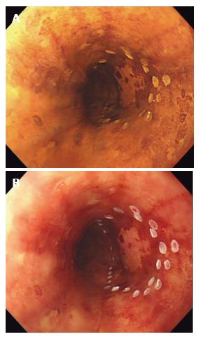
Figure 1 Chromoendoscopy with Lugol’s iodine.
A: Unstained area seen within the marking after spraying diluted Lugol’s solution with spray catheter; B: After observing for several minutes, the unstained area turned into pink color, suggesting HGIN and squamous cell carcinoma.
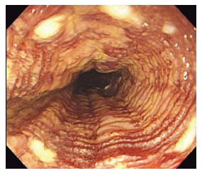
Figure 2 “Tatami sign” is commonly seen after iodine staining.
It is characterized by regular, fine circular folds of the Lugol’s unstained area. This is typically seen when lesions are confined to the muscularis mucosal.
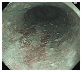
Figure 3 Brownish areas seen under non-magnifying narrow-band imaging can be seen with inflammation, low-grade and high-grade intraepithelial neoplasm, and squamous cell carcinoma.
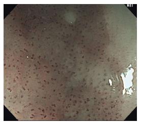
Figure 4 Abnormal intraepithelial papillary capillary loop, a superficial fine vascular network of the esophageal mucosa.
Type B1 vessels, shown here, are identified as dot-like microvessels under non-magnifying narrow-band imaging or low magnified ME-NBI.
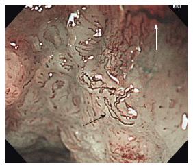
Figure 5 Abnormal intraepithelial papillary capillary loop.
Type B2 (black arrow) can be recognized as stretched and markedly elongated microvessels vs type B3 (white arrow) microvessels which are highly dilated, abnormal vessels.
- Citation: Shimamura Y, Ikeya T, Marcon N, Mosko JD. Endoscopic diagnosis and treatment of early esophageal squamous neoplasia. World J Gastrointest Endosc 2017; 9(9): 438-447
- URL: https://www.wjgnet.com/1948-5190/full/v9/i9/438.htm
- DOI: https://dx.doi.org/10.4253/wjge.v9.i9.438


