Copyright
©The Author(s) 2017.
World J Gastrointest Endosc. Sep 16, 2017; 9(9): 438-447
Published online Sep 16, 2017. doi: 10.4253/wjge.v9.i9.438
Published online Sep 16, 2017. doi: 10.4253/wjge.v9.i9.438
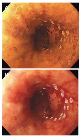
Figure 1 Chromoendoscopy with Lugol’s iodine.
A: Unstained area seen within the marking after spraying diluted Lugol’s solution with spray catheter; B: After observing for several minutes, the unstained area turned into pink color, suggesting HGIN and squamous cell carcinoma.
- Citation: Shimamura Y, Ikeya T, Marcon N, Mosko JD. Endoscopic diagnosis and treatment of early esophageal squamous neoplasia. World J Gastrointest Endosc 2017; 9(9): 438-447
- URL: https://www.wjgnet.com/1948-5190/full/v9/i9/438.htm
- DOI: https://dx.doi.org/10.4253/wjge.v9.i9.438


