Copyright
©The Author(s) 2017.
World J Gastrointest Endosc. Mar 16, 2017; 9(3): 145-148
Published online Mar 16, 2017. doi: 10.4253/wjge.v9.i3.145
Published online Mar 16, 2017. doi: 10.4253/wjge.v9.i3.145
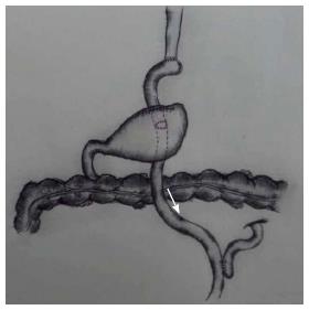
Figure 1 Modified double tracks anastomosis.
The arrow indicates the long limb.
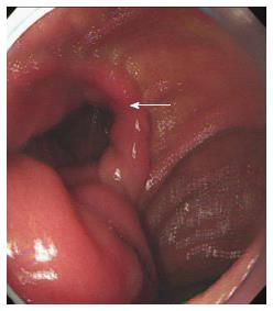
Figure 2 Gastroscopy showing another anastomosis (arrow, gastrojejunal anastomosis) after the esophagojejunal anastomosis.
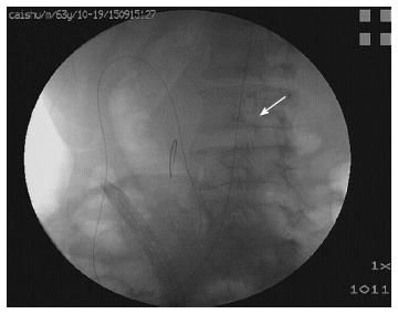
Figure 3 Guide-wire (arrow) placed for introducingce the duodenoscope.
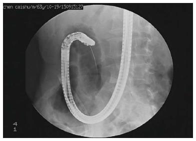
Figure 4 Duodenoscope introduced by the guide wire and arriving at the major papilla.
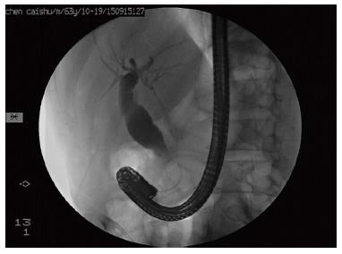
Figure 5 Cholangiogram of endoscopic retrograde cholangiopancreat-ography demonstrating dilated common bile duct with a filling defect.
- Citation: Wang XS, Wang F, Li QP, Miao L, Zhang XH. Endoscopic retrograde cholangiopancretography in modified double tracks anastomosis with anastomotic stenosis. World J Gastrointest Endosc 2017; 9(3): 145-148
- URL: https://www.wjgnet.com/1948-5190/full/v9/i3/145.htm
- DOI: https://dx.doi.org/10.4253/wjge.v9.i3.145


