Copyright
©The Author(s) 2016.
World J Gastrointest Endosc. Feb 25, 2016; 8(4): 244-251
Published online Feb 25, 2016. doi: 10.4253/wjge.v8.i4.244
Published online Feb 25, 2016. doi: 10.4253/wjge.v8.i4.244
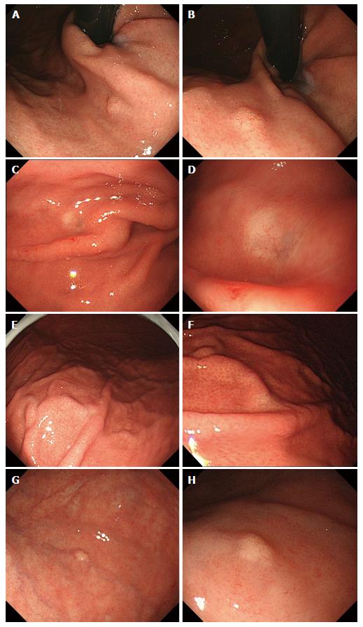
Figure 1 Endoscopic findings: Case 1 (A, B), case 2 (C, D), case 3 (E, F) and case 4 (G, H).
Conventional endoscopy on white-light imaging showed flat lesions with whitish discoloration (C-F) or yellowish submucosal tumor shapes (A, B, G, H). Dilated vessels were shown on the surface of tumors (B, D, F, H).
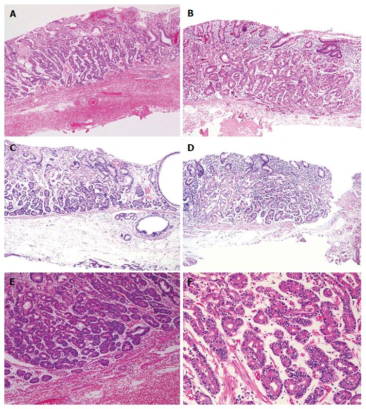
Figure 2 Histological findings (hematoxylin and eosin staining): Case 1 (A, E, F), case 2 (B), case 3 (C) and case 4 (D).
Well-differentiated adenocarcinomas with columnar cells that mimicked fundic gland cells were observed. Tumors histologically arose in the deeper zone of gastric mucosa (A-D). Irregularly anastomosing glandular structures with mildly enlarged and hyperchromatic nuclei were observed (E, F). Minimum carcinoma invasion of the submucosal layer was detected (C, D). Neither lymphatic nor vascular invasion was observed. Magnification: A-D (low-power view × 100), E (high-power view × 200), F (high-power view × 400).
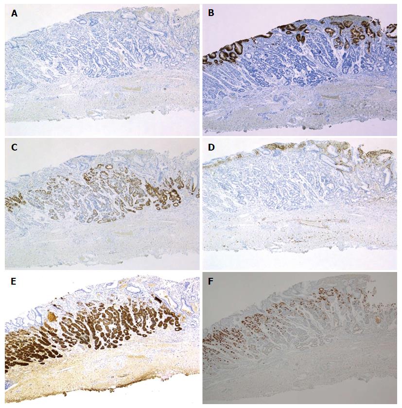
Figure 3 Immunohistochemical staining: Case 1.
Tumor cells were diffusely positive for MUC6 (C) and pepsinogen-I (E), partially positive for H+/K+-ATPase in scattered locations around the tumor margin (F), negative staining for MUC2 (A), MUC5AC (B) and CD10 (D). Magnification: A-F (low-power view × 40). MUC: Mucin.
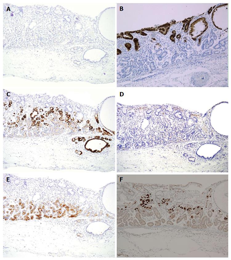
Figure 4 Immunohistochemical staining: Case 3.
Tumor cells were diffusely positive for MUC6 (C) and pepsinogen-I (E), partially positive for H+/K+-ATPase in scattered locations around the tumor margin (F), negative staining for MUC2 (A), MUC5AC (B) and CD10 (D). Magnification: A-F (low-power view × 100). MUC: Mucin.
- Citation: Tohda G, Osawa T, Asada Y, Dochin M, Terahata S. Gastric adenocarcinoma of fundic gland type: Endoscopic and clinicopathological features. World J Gastrointest Endosc 2016; 8(4): 244-251
- URL: https://www.wjgnet.com/1948-5190/full/v8/i4/244.htm
- DOI: https://dx.doi.org/10.4253/wjge.v8.i4.244


