Copyright
©The Author(s) 2016.
World J Gastrointest Endosc. Sep 16, 2016; 8(17): 572-583
Published online Sep 16, 2016. doi: 10.4253/wjge.v8.i17.572
Published online Sep 16, 2016. doi: 10.4253/wjge.v8.i17.572
Figure 1 Lesions compatible with Crohn's disease by capsule endoscopy.
A: Edema; B: Ulcers; C: Strictures.
Figure 2 Aphthous erosions detected by capsule endoscopy.
A: Aphtha; B: Surface erosion; C: Aphthoid erosions. The capsule may detect superficial intestinal lesions in a patient with Crohn’s disease that are overlooked by radiographic techniques and inaccessible to ileocolonoscopy.
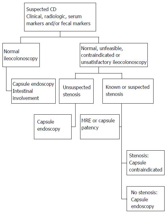
Figure 3 A diagnostic protocol for suspected Crohn’s disease[127].
When CD is suspected ileocolonoscopy should be the first study to be performed, with capsule endoscopy ensuing when results are normal, unsatisfactory or not are achieved Ileoscopy. If intestinal stenosis is suspected, a test capsule should be used to confirm the feasibility of capsule endoscopy. CD: Crohn's disease; MRE: Enterography with nuclear magnetic resonance.
- Citation: Luján-Sanchis M, Sanchis-Artero L, Larrey-Ruiz L, Peño-Muñoz L, Núñez-Martínez P, Castillo-López G, González-González L, Clemente CB, Albert Antequera C, Durá-Ayet A, Sempere-Garcia-Argüelles J. Current role of capsule endoscopy in Crohn’s disease. World J Gastrointest Endosc 2016; 8(17): 572-583
- URL: https://www.wjgnet.com/1948-5190/full/v8/i17/572.htm
- DOI: https://dx.doi.org/10.4253/wjge.v8.i17.572


