Copyright
©The Author(s) 2016.
World J Gastrointest Endosc. Aug 10, 2016; 8(15): 533-540
Published online Aug 10, 2016. doi: 10.4253/wjge.v8.i15.533
Published online Aug 10, 2016. doi: 10.4253/wjge.v8.i15.533
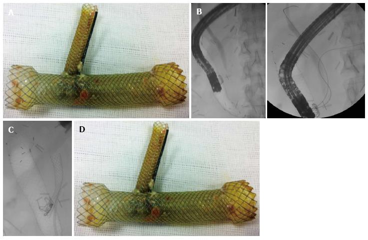
Figure 1 Fluoroscopy.
A: Trans-stent duodenoscopy. Cholangiopancreatography already performed prior to enteral stenting; B: Two guidewires (biliary and pancreatic) inside the biliary and the pancreatic ducts; C: Final fluoroscopic image of a 6 cm × 10 mm biliary SEMS and a 7 Fr × 7 cm plastic pancreatic stent draining inside the enteral stent; D: After 4 wk, the multistent complex was removed endoscopically.
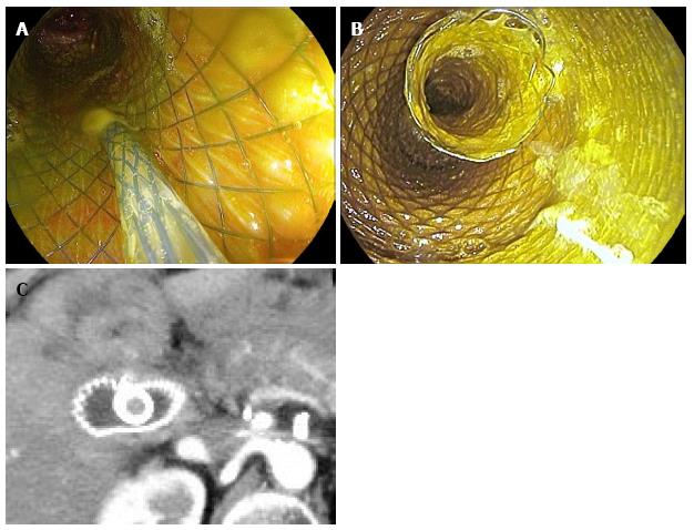
Figure 2 Bilio-enteric external fistula after pancreaticoduodenectomy.
A: An 8 mm × 4 cm biliary SEMS was inserted into the common bile duct through the meshes of the enteral SEMS; B: Final stent complex at the end of the procedure; C: Detail of CT scan performed two days after the intervention showing the relationship between the two SEMS (biliary stent protruding inside the enteral).
- Citation: Mutignani M, Dioscoridi L, Dokas S, Aseni P, Carnevali P, Forti E, Manta R, Sica M, Tringali A, Pugliese F. Endoscopic multiple metal stenting for the treatment of enteral leaks near the biliary orifice: A novel effective rescue procedure. World J Gastrointest Endosc 2016; 8(15): 533-540
- URL: https://www.wjgnet.com/1948-5190/full/v8/i15/533.htm
- DOI: https://dx.doi.org/10.4253/wjge.v8.i15.533


