Copyright
©The Author(s) 2016.
World J Gastrointest Endosc. Jun 25, 2016; 8(12): 444-450
Published online Jun 25, 2016. doi: 10.4253/wjge.v8.i12.444
Published online Jun 25, 2016. doi: 10.4253/wjge.v8.i12.444
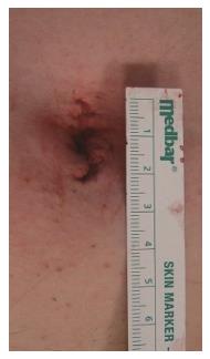
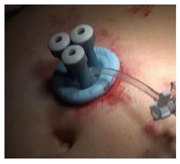
Figure 1 A 2-cm umbilical single-port incision.
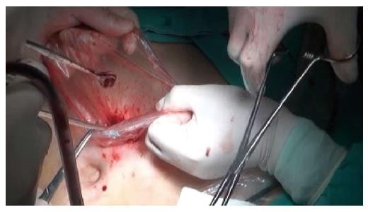
Figure 2 Fragmentation of the specimen without extension of the single-port incision.
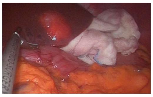
Figure 3 A peripherally located benign lesion.
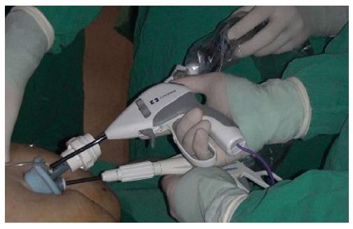
Figure 4 Conflict between the surgeon and the camera holder extracorporeally.
Figure 5 The entry of the port should be selected based on the patient’s body type and the location of the lesion.
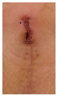
Figure 6 A single-port laparoscopic liver resection incision a patient with a previous history of colon resection.
- Citation: Karabicak I, Karabulut K. Single port laparoscopic liver surgery: A minireview. World J Gastrointest Endosc 2016; 8(12): 444-450
- URL: https://www.wjgnet.com/1948-5190/full/v8/i12/444.htm
- DOI: https://dx.doi.org/10.4253/wjge.v8.i12.444


