Copyright
©The Author(s) 2015.
World J Gastrointest Endosc. Apr 16, 2015; 7(4): 381-388
Published online Apr 16, 2015. doi: 10.4253/wjge.v7.i4.381
Published online Apr 16, 2015. doi: 10.4253/wjge.v7.i4.381
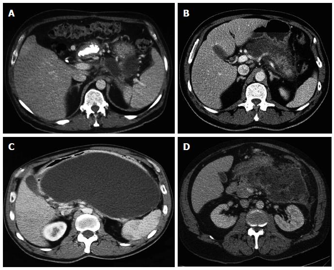
Figure 1 Acute peripancreatic fluid collection (A), acute necrotic collection (B), pancreatic pseudocyst (C), and walled-of necrosis (D).
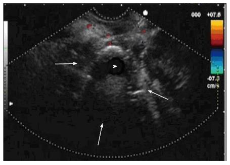
Figure 2 Endoscopic ultrasonography image of walled-off necrosis collection.
The limits of the walled-off necrosis are signalled by the arrows. The necrotic content is marked with arrowhead.
Figure 3 Pancreatic pseudocyst (A), endoscopic dilation of transmural tract (B), and three double-pigtail plastic stents placed (C).
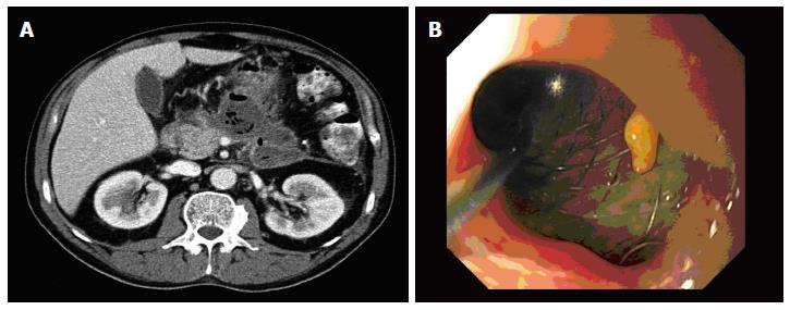
Figure 4 Walled-of necrosis with air content suspicious of fistulization or infection (A) and internal migration of stent (B).
- Citation: Ruiz-Clavijo D, Higuera BGL, Vila JJ. Advances in the endoscopic management of pancreatic collections. World J Gastrointest Endosc 2015; 7(4): 381-388
- URL: https://www.wjgnet.com/1948-5190/full/v7/i4/381.htm
- DOI: https://dx.doi.org/10.4253/wjge.v7.i4.381


