Copyright
©The Author(s) 2015.
World J Gastrointest Endosc. Oct 10, 2015; 7(14): 1150-1156
Published online Oct 10, 2015. doi: 10.4253/wjge.v7.i14.1150
Published online Oct 10, 2015. doi: 10.4253/wjge.v7.i14.1150
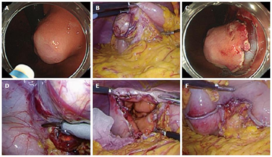
Figure 1 Laparoscopic endoscopic cooperative surgery for gastric submucosal tumor.
A: The tumor is located in the lesser curvature of the middle third of the stomach; B: The stomach was mobilized by dividing the gastrocolic omentum and the lesser curvature vessels near the tumor by laparoscopic dissection; C: A circumferential incision was made around the tumor by an endoscopic submucosal dissection technique using an insulation-tipped diathermic electrosurgical knife; D: The seromuscular layer of the stomach was dissected along the incision line using the laparosonic coagulating shears; E, F: The post-excisional hole in the stomach was closed using a laparoscopic linear stapling device.
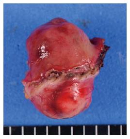
Figure 2 Gross appearance of the resected specimen.
The tumor is a mixed-type, with a predominant intragastric component. The resection margin of healthy gastric wall is limited to the minimum necessary. The pathological diagnosis confirmed a gastrointestinal stromal tumor, classified as low risk.
- Citation: Namikawa T, Hanazaki K. Laparoscopic endoscopic cooperative surgery as a minimally invasive treatment for gastric submucosal tumor. World J Gastrointest Endosc 2015; 7(14): 1150-1156
- URL: https://www.wjgnet.com/1948-5190/full/v7/i14/1150.htm
- DOI: https://dx.doi.org/10.4253/wjge.v7.i14.1150


