Copyright
©The Author(s) 2015.
World J Gastrointest Endosc. Oct 10, 2015; 7(14): 1142-1149
Published online Oct 10, 2015. doi: 10.4253/wjge.v7.i14.1142
Published online Oct 10, 2015. doi: 10.4253/wjge.v7.i14.1142
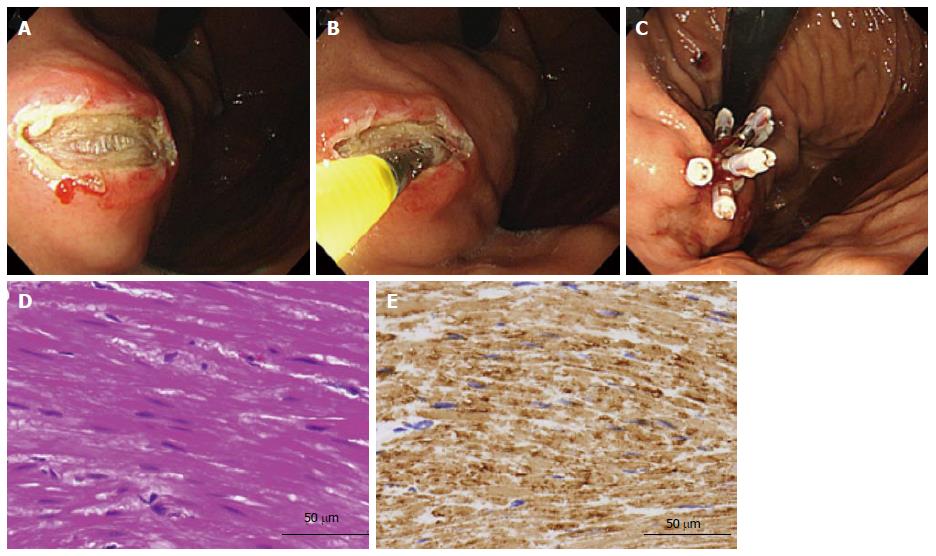
Figure 1 Mucosal cutting biopsy of a submucosal tumor with intraluminal growth in the lesser curvature of the corpus.
A: The mucosal incision was made by a needle-knife after an injection of saline; B: Biopsy specimen obtained from the tumor using biopsy forceps under direct observation; C: The incision was closed with hemoclips; D: Histological examination of the biopsied specimen showing a spindle cell without mitotic figures (HE); E: Immunohistochemical staining was positive for desmin. This lesion was diagnosed histologically as a leiomyoma.
Figure 2 Endoscopic ultrasonography-guided fine-needle aspiration biopsy of a gastrointestinal stromal tumor with extraluminal growth.
A: EUS-FNAB of a hypoechoic lesion in the muscularis propria layer showing extraluminal growth; B: Histological finding showing spindle cells in the EUS-FNAB specimen (HE); C: Immunohistochemical staining is positive for c-kit. EUS-FNAB: Endoscopic ultrasonography-guided fine-needle aspiration biopsy.
- Citation: Ikehara H, Li Z, Watari J, Taki M, Ogawa T, Yamasaki T, Kondo T, Toyoshima F, Kono T, Tozawa K, Ohda Y, Tomita T, Oshima T, Fukui H, Matsuda I, Hirota S, Miwa H. Histological diagnosis of gastric submucosal tumors: A pilot study of endoscopic ultrasonography-guided fine-needle aspiration biopsy vs mucosal cutting biopsy. World J Gastrointest Endosc 2015; 7(14): 1142-1149
- URL: https://www.wjgnet.com/1948-5190/full/v7/i14/1142.htm
- DOI: https://dx.doi.org/10.4253/wjge.v7.i14.1142


