Copyright
©2014 Baishideng Publishing Group Inc.
World J Gastrointest Endosc. Nov 16, 2014; 6(11): 568-570
Published online Nov 16, 2014. doi: 10.4253/wjge.v6.i11.568
Published online Nov 16, 2014. doi: 10.4253/wjge.v6.i11.568
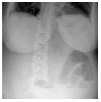
Figure 1 Plane abdominal X-ray demonstrated the findings of intestinal obstruction.
The small bowel segment was dilated and there were air-fluid levels.
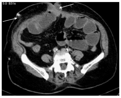
Figure 2 Incarcerated intestinal loops in the umbilical trocar site.
Arrows point out the abdominal drain and the incarcerated intestinal loops through the umbilicus.
- Citation: Sumer F, Kayaalp C, Yagci MA, Otan E, Kocaaslan H. Early endoscopic retrograde cholangiopancreatography after laparoscopic cholecystectomy can strain the occurrence of trocar site hernia. World J Gastrointest Endosc 2014; 6(11): 568-570
- URL: https://www.wjgnet.com/1948-5190/full/v6/i11/568.htm
- DOI: https://dx.doi.org/10.4253/wjge.v6.i11.568


