Copyright
©2013 Baishideng Publishing Group Co.
World J Gastrointest Endosc. Sep 16, 2013; 5(9): 465-467
Published online Sep 16, 2013. doi: 10.4253/wjge.v5.i9.465
Published online Sep 16, 2013. doi: 10.4253/wjge.v5.i9.465
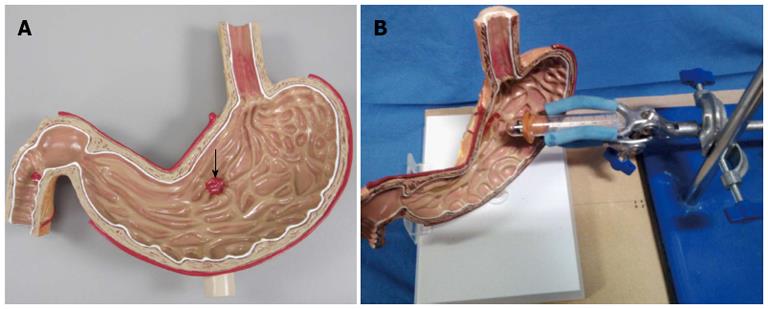
Figure 1 Phantom model (A) and task simulator setting (B).
A: The arrow points to the gastric ulcer (‘‘1/2 diameter and 3/16” depth).
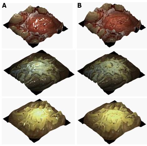
Figure 2 Three-dimensional representation of images captured for the 3 models: red, white and yellow.
A: Original three-dimensional (3-D) represented images; B: The processed 3-D represented images using the highlight suppression algorithm.
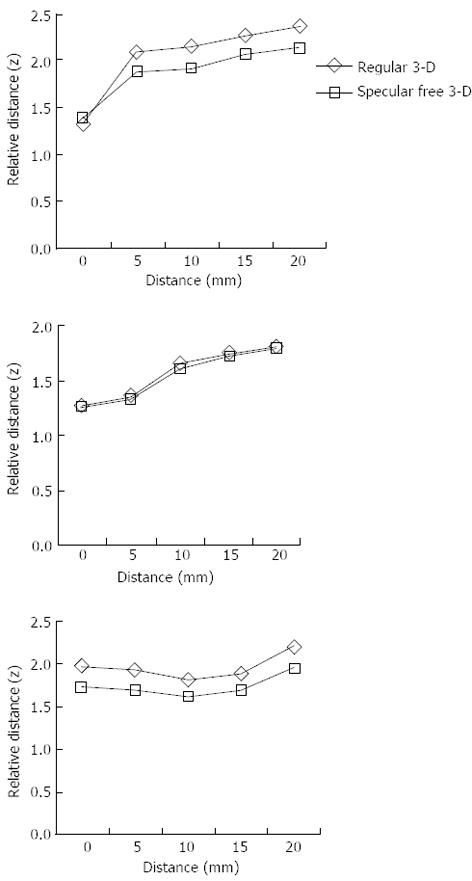
Figure 3 Relative distance of three-dimensional representation calculated over images taken from various distances of the capsule from the models.
- Citation: Koulaouzidis A, Karargyris A. Use of enhancement algorithm to suppress reflections in 3-D reconstructed capsule endoscopy images. World J Gastrointest Endosc 2013; 5(9): 465-467
- URL: https://www.wjgnet.com/1948-5190/full/v5/i9/465.htm
- DOI: https://dx.doi.org/10.4253/wjge.v5.i9.465


