Copyright
©2013 Baishideng Publishing Group Co.
World J Gastrointest Endosc. Sep 16, 2013; 5(9): 440-445
Published online Sep 16, 2013. doi: 10.4253/wjge.v5.i9.440
Published online Sep 16, 2013. doi: 10.4253/wjge.v5.i9.440
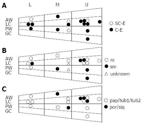
Figure 1 Locations of 17 false-negative gastric cancers.
A: Open circles indicate the false-negative gastric cancer (FN-GC) lesions found by small-caliber endoscope (SC-E), and closed circles indicate the FN-GCs found by conventional endoscopy (C-E); B: Open circles indicate FN-GC lesions that invaded to the mucosal layer and closed circles indicate the FN-GC lesions that invaded to the submucosal layer. Open triangles indicate lesions with unknown depth; C: Open circles indicate the FN-GC lesions of pathologically differentiated types (papillary adenocarcinoma, well or moderately differentiated adenocarcinoma) and closed circles indicate the FN-GC lesions of pathologically diffuse types (poorly differentiated adenocarcinoma or signet ring cell carcinoma). m: Mucosal layer; sm: Submucosal layer; pap: Papillary adenocarcinoma; tub1: Well differentiated tubular adenocarcinoma; tub2: Moderately differentiated adenocarcinoma; por: Poorly differentiated adenocarcinoma; sig: Signet ring cell carcinoma; AW: Anterior wall; PW: Posterior wall; LC: Lesser curvature; GC: Greater curvature; U: Upper; M: Middle; L: Lower.
- Citation: Kataoka H, Mizuno K, Hayashi N, Tanaka M, Nishiwaki H, Ebi M, Mizoshita T, Mori Y, Kubota E, Tanida S, Kamiya T, Joh T. Diagnostic utility of small-caliber and conventional endoscopes for gastric cancer and analysis of endoscopic false-negative gastric cancers. World J Gastrointest Endosc 2013; 5(9): 440-445
- URL: https://www.wjgnet.com/1948-5190/full/v5/i9/440.htm
- DOI: https://dx.doi.org/10.4253/wjge.v5.i9.440


