Copyright
©2013 Baishideng Publishing Group Co.
World J Gastrointest Endosc. Aug 16, 2013; 5(8): 402-406
Published online Aug 16, 2013. doi: 10.4253/wjge.v5.i8.402
Published online Aug 16, 2013. doi: 10.4253/wjge.v5.i8.402
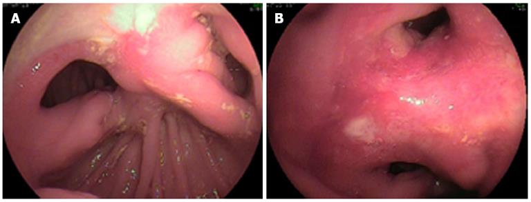
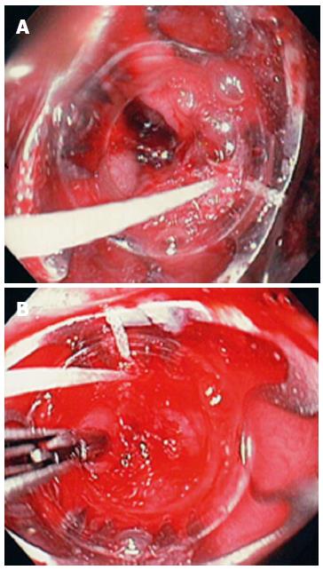
Figure 1 Billroth II anatomy.
A clean-based ulcer is present at the anastomosis (A), the lumen to both the afferent and efferent limbs is patent (A); the gastrocolic fistula opening was located at the upper end of the anastomosis (B).
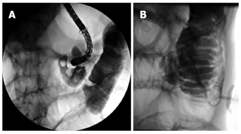
Figure 2 Insertion of the scope through this orifice lead to the colon (A), after placing the jejunostomy, water soluble contrast was injected confirming its perfect intra-jejunal position (B).
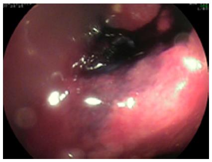
Figure 3 Before closing the fistula the entrance into the orifice was marked with India ink.
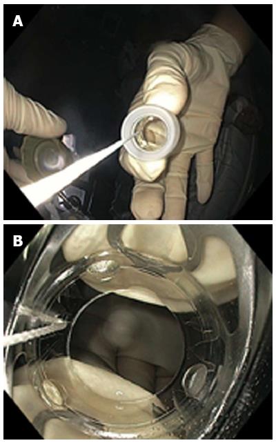
Figure 4 T the over-the-scope-clip -system come loaded onto a transparent cap which is attached to the tip of the scope.
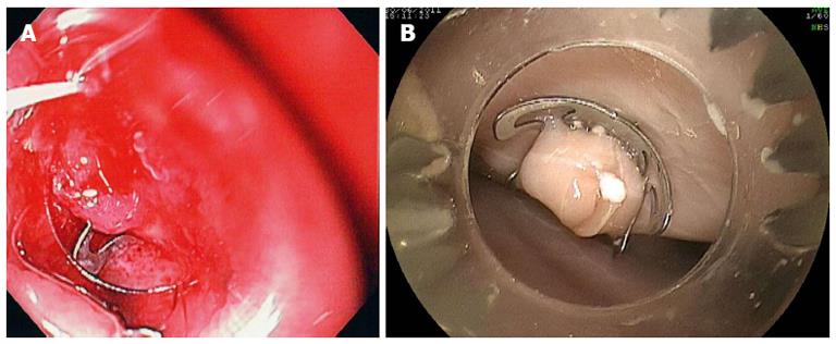
Figure 5 The over-the-scope-clip-system was approximated to the fistula (A) and the proximal edge of the fistula was pulled inside the transparent cap of the over-the-scope-clip-system using the Twin Grasper (B).
Figure 6 Once enough tissue was present inside the cap the over-the-scope-clip device was released.
(A) Example of deployed over-the-scope-clip -system in experimental perforation in an ex-vivo pig stomach (B).
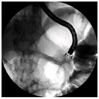
Figure 7 Radiologic view of the over-the-scope-clip -system (‘‘bear trap”).
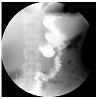
Figure 8 Barium study documents complete closure of the fistula.
The contrast flows into the jejunal limbs.
- Citation: Mönkemüller K, Peter S, Alkurdi B, Ramesh J, Popa D, Wilcox CM. Endoscopic closure of a gastrocolic fistula using the over-the-scope-clip-system. World J Gastrointest Endosc 2013; 5(8): 402-406
- URL: https://www.wjgnet.com/1948-5190/full/v5/i8/402.htm
- DOI: https://dx.doi.org/10.4253/wjge.v5.i8.402


