Copyright
©2013 Baishideng Publishing Group Co.
World J Gastrointest Endosc. Apr 16, 2013; 5(4): 180-185
Published online Apr 16, 2013. doi: 10.4253/wjge.v5.i4.180
Published online Apr 16, 2013. doi: 10.4253/wjge.v5.i4.180
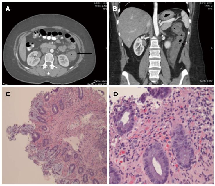
Figure 1 Computed tomography scan and histopathology.
A, B: Computed tomography scan shows thickening of the colonic wall involving the descending colon (arrows); C, D: Histopathology shows: the overlying surface mucosa is eroded, the lamina propria is partially hyalinized with fibropurulent exudate and acute inflammation, consistent with ischemic colitis.
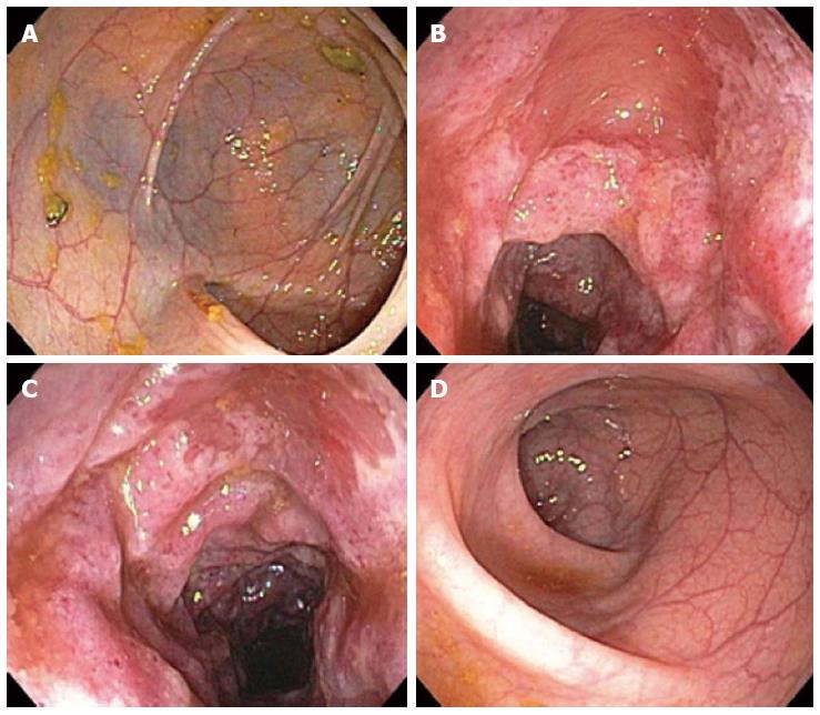
Figure 2 Colonoscopy shows.
A: Normal mucosa of the right colon (hepatic flexure); B, C: Erythematous, edematous, erosive, and ulcerated mucosa of the splenic flexure of the colon, consistent with ischemic colitis; D: Normal mucosa of the sigmoid colon.
- Citation: Sherid M, Samo S, Sulaiman S, Gaziano JH. Ischemic colitis induced by the newly reformulated multicomponent weight-loss supplement Hydroxycut®. World J Gastrointest Endosc 2013; 5(4): 180-185
- URL: https://www.wjgnet.com/1948-5190/full/v5/i4/180.htm
- DOI: https://dx.doi.org/10.4253/wjge.v5.i4.180


