Copyright
©2012 Baishideng.
World J Gastrointest Endosc. Sep 16, 2012; 4(9): 434-437
Published online Sep 16, 2012. doi: 10.4253/wjge.v4.i9.434
Published online Sep 16, 2012. doi: 10.4253/wjge.v4.i9.434
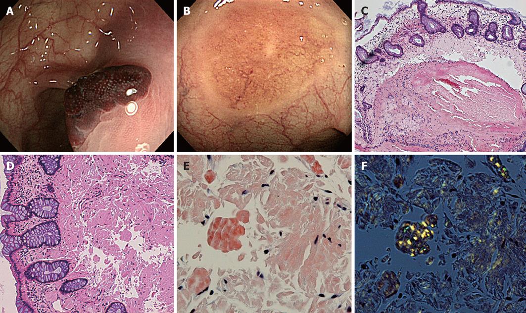
Figure 1 Initial colonoscopy revealed reddish elevated lesions within plaque-like erythema in the sigmoid and transverse colon.
A: Initial colonoscopy showing a submucosal hematoma in the transverse colon; B: Repeat colonoscopy showing a discoid discoloration where the submucosal hematoma was located; C: Biopsy from submucosal hematoma showing mucosal hemorrhages and deposition of amorphous material in the lamina propria (hematoxylin-eosin stain); D: Biopsy from discoid lesion showing deposition of amorphous material in the lamina propria and in the vessel walls of the submucosa (hematoxylin-eosin stain); E: Congo red stain showing amyloid deposition; F: Apple-green birefringence under polarized light.
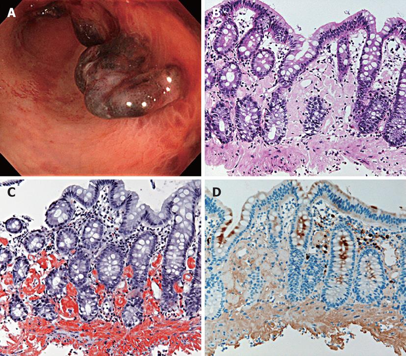
Figure 2 Endoscopic biopsies were performed for the submucosal hematomas.
A: Colonoscopy showing submucosal hematomas from the ascending colon to the rectum; B and C: Biopsy showing mucosal hemorrhages and deposition of amorphous material in the lamina propria and in the vessel walls of the submucosa (hematoxylin-eosin stain and direct fast scarlet stain); D: Staining with κ was positive.
- Citation: Yoshii S, Mabe K, Nosho K, Yamamoto H, Yasui H, Okuda H, Suzuki A, Fujita M, Sato T. Submucosal hematoma is a highly suggestive finding for amyloid light-chain amyloidosis: Two case reports. World J Gastrointest Endosc 2012; 4(9): 434-437
- URL: https://www.wjgnet.com/1948-5190/full/v4/i9/434.htm
- DOI: https://dx.doi.org/10.4253/wjge.v4.i9.434


