Copyright
©2012 Baishideng.
World J Gastrointest Endosc. Aug 16, 2012; 4(8): 362-367
Published online Aug 16, 2012. doi: 10.4253/wjge.v4.i8.362
Published online Aug 16, 2012. doi: 10.4253/wjge.v4.i8.362
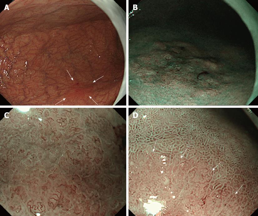
Figure 1 Narrow band imaging findings.
A: Ordinary [non magnifying endoscopy with narrow band imaging (ME-NBI)] endoscopic findings of intramucosal gastric carcinoma. A flare-like flat-type (0-IIb) lesion measuring 25 mm in diameter, with an unclear border, was observed at the greater curvature of the inferior gastric body (arrows); B: NBI finding of the lesion (non-ME); C: Strongly-magnified NBI finding at the lesion center; D: Moderately-magnified NBI finding at the margin: a demarcation line between the cancerous and non-cancerous regions could be recognized based on differences in the fine structure of mucosa and microvascular features (arrows). This was established as an imaginary boundary.
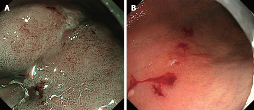
Figure 2 Biopsy findings and standard observation biopsy.
A: As shown in the photograph, biopsy was performed by measuring a distance of approximately 1.8 mm using forceps (tip diameter: 1.8 mm), regarding two regions sandwiching the boundary estimated on NBI as cancerous and non-cancerous; B: Standard observation after biopsy.
- Citation: Nonaka K, Namoto M, Kitada H, Shimizu M, Ochiai Y, Togawa O, Nakao M, Nishimura M, Ishikawa K, Arai S, Kita H. Usefulness of the DL in ME with NBI for determining the expanded area of early-stage differentiated gastric carcinoma. World J Gastrointest Endosc 2012; 4(8): 362-367
- URL: https://www.wjgnet.com/1948-5190/full/v4/i8/362.htm
- DOI: https://dx.doi.org/10.4253/wjge.v4.i8.362


