Copyright
©2012 Baishideng Publishing Group Co.
World J Gastrointest Endosc. Jul 16, 2012; 4(7): 269-280
Published online Jul 16, 2012. doi: 10.4253/wjge.v4.i7.269
Published online Jul 16, 2012. doi: 10.4253/wjge.v4.i7.269
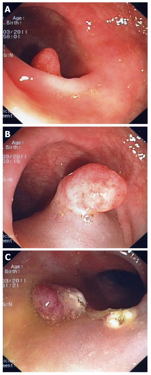
Figure 1 Difficult polyp located “behind” a fold (A).
A closer look indicates that this is a large sessile polyp (B). The polyp was resected using mucosectomy technique (C).
Figure 2 Large polyp located in the transverse colon (A).
A closer inspection reveals that this polyp has a thick stalk (B); After injecting the stalk with adrenaline-saline mixture the snare was placed around it; Notice that the snare exits the scope at 5 o’clock position (C); A clip was placed at the base of the stalk to prevent post-polypectomy bleeding (D).
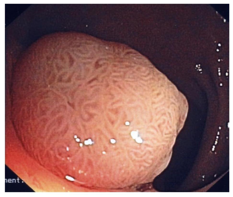
Figure 3 Large sessile polyp with cerebriform pit-pattern.
This polyp was an adenoma with high-grade dysplasia.
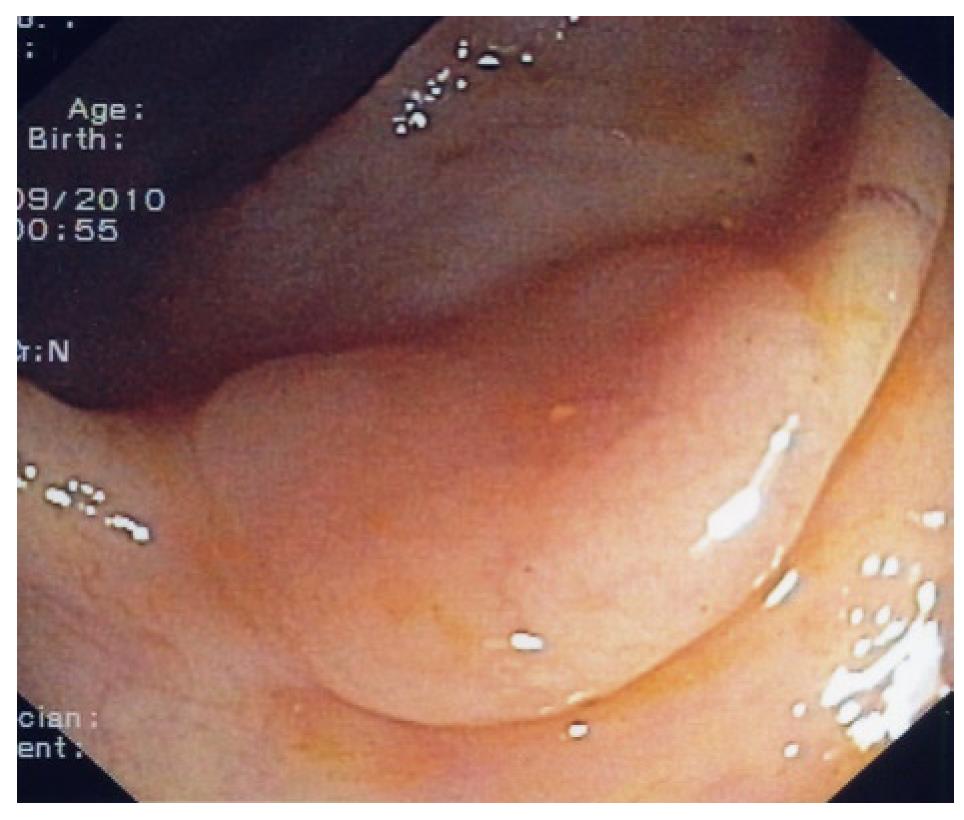
Figure 4 Typical flat polyp located on top of a fold.
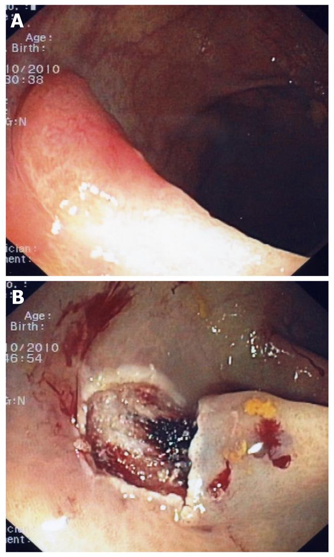
Figure 5 Flat polyp with irregular vascular pattern (A).
Mucosectomy site (B); This polyp contained invasive cancer. A subsequent laparoscopic segmental section of the colon was performed.
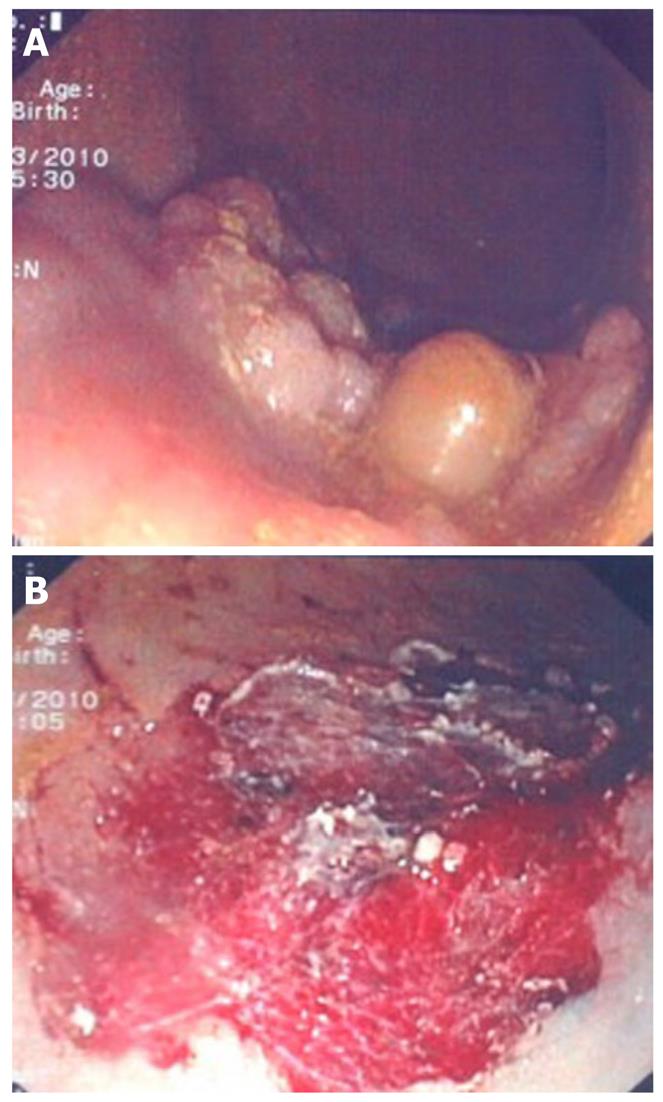
Figure 6 Large sessile polyp (A) and polyp site after performing piece-meal mucosectomy (B).
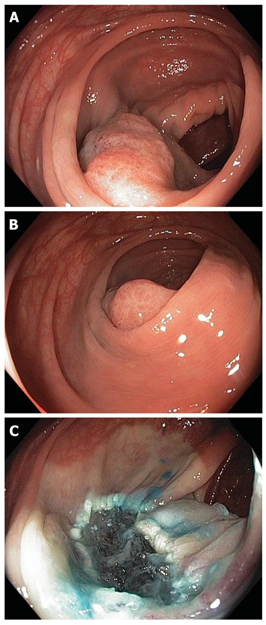
Figure 7 Sessile polyp located behind a fold (A), the creation of the submucosal cushion enabled the clear visualization of the polyp (B) and endoscopic resection site (C).
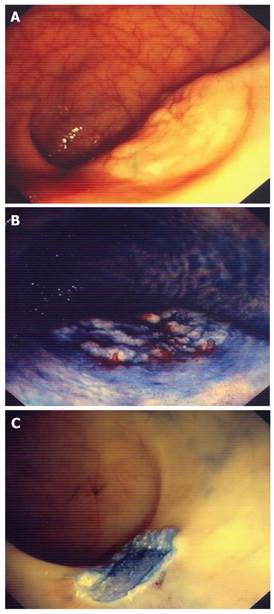
Figure 8 Flat polyp located in the rectum (A), demarcation of the polyp edges and surface with standard chromoendoscopy (indigo carmine) (B) and endoscopic mucosal resection site (C).
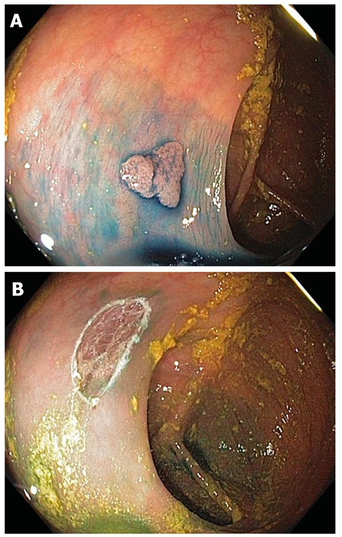
Figure 9 Sessile polyp located in the transverse colon.
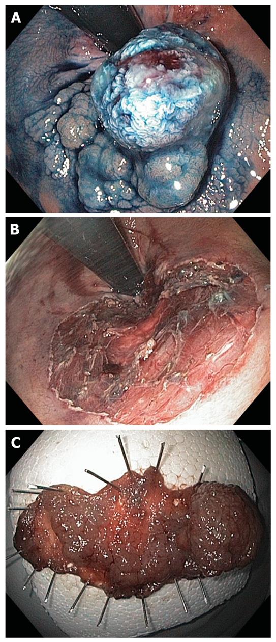
Demarcation of the polyp margins and surface with standard chromoendoscopy (methylene blue) (A); Endoscopic mucosal resection site (B).
Figure 10 Giant rectal polyp (A), the polyp was resected using endoscopic submucosal dissection-technique (B) and an en-bloc resection was possible (C).
- Citation: Vormbrock K, Mönkemüller K. Difficult colon polypectomy. World J Gastrointest Endosc 2012; 4(7): 269-280
- URL: https://www.wjgnet.com/1948-5190/full/v4/i7/269.htm
- DOI: https://dx.doi.org/10.4253/wjge.v4.i7.269


