
















Copyright: ©Author(s) 2026.
World J Gastrointest Endosc. Sep 16, 2026; 18(9): 124149
Published online Sep 16, 2026. doi: 10.4253/wjge.124149
Published online Sep 16, 2026. doi: 10.4253/wjge.124149
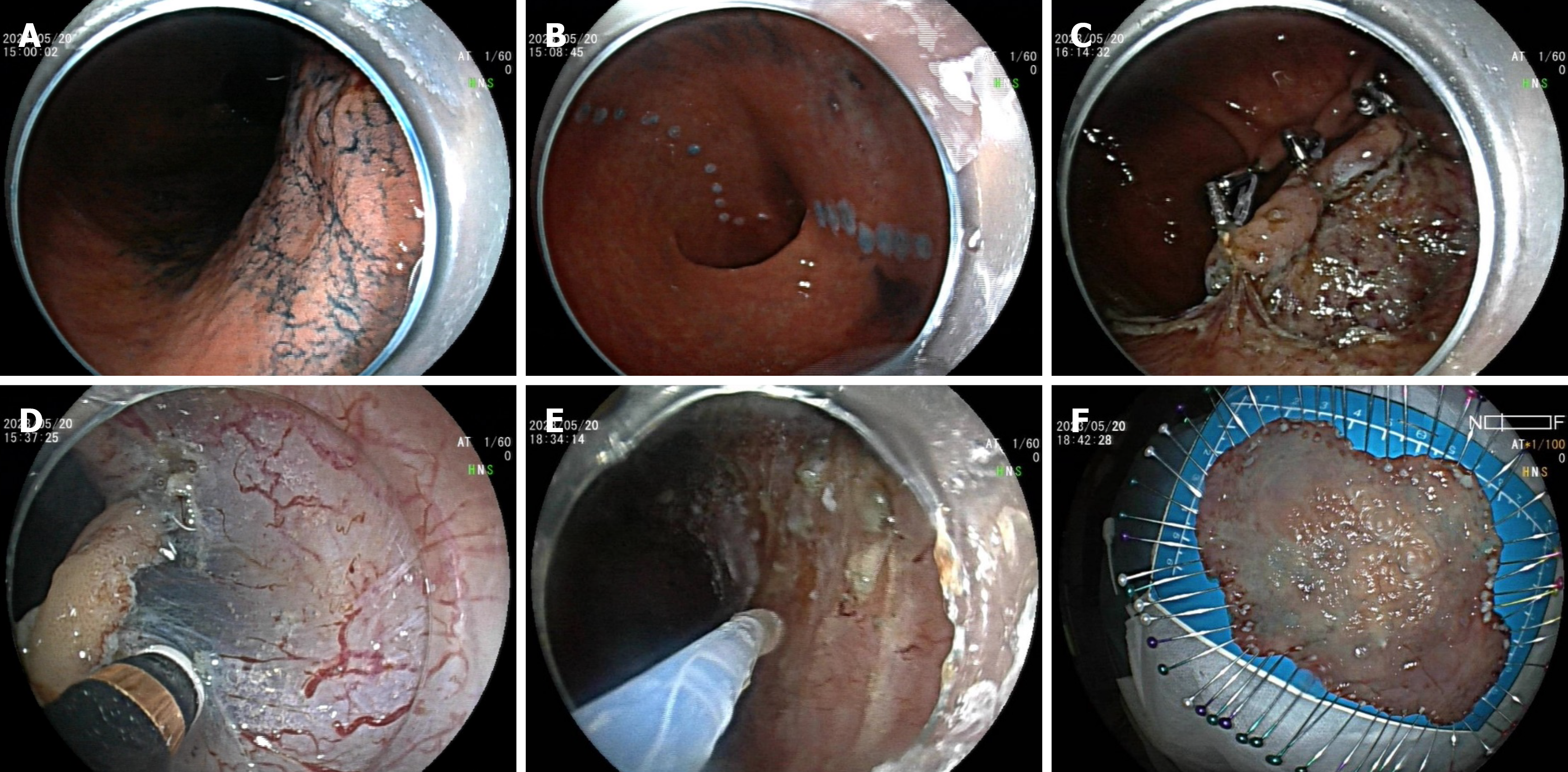
Figure 1 Endoscopy images during endoscopic submucosal dissection.
A: After spraying indigo rouge, the stain on the lesion is obvious and the edge of the lesion is clear; B: Intraoperative marking showed that the lesion extended from the posterior wall and lesser curvature of the lower gastric body to the gastric angulus and gastric antrum; C and D: Under the traction and assistance of titanium clips, the submucosal lesion was completely removed by endoscopic submucosal dissection; E: Spraying tissue adhesive onto the surface of the resection defect to prevent postoperative bleeding; F: Complete resection of the lesion, about 60 mm × 100 mm in size.
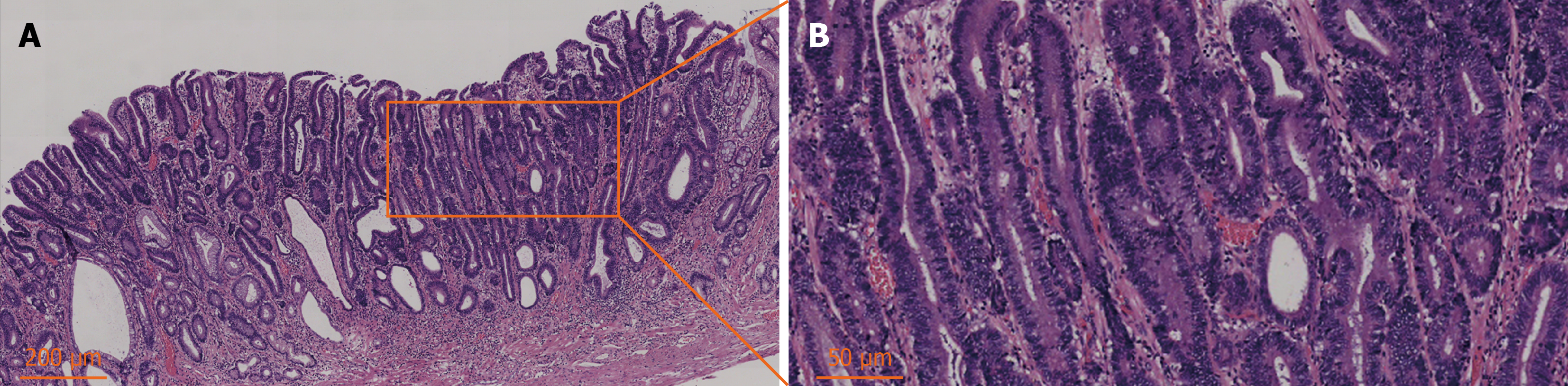
Figure 2 Findings of histopathology examination of the endoscopically resected specimen.
A and B: There were no lesions at the lateral and basal incisal margins of the lesion. High-grade intraepithelial neoplasia presented a multifocal and scattered distribution, some of which showed adenocarcinoma in situ-like changes without invading the stroma and muscularis mucosa. The surrounding mucosa presented low-grade intraepithelial neoplasia, showing intestinal metaplasia and atrophy.
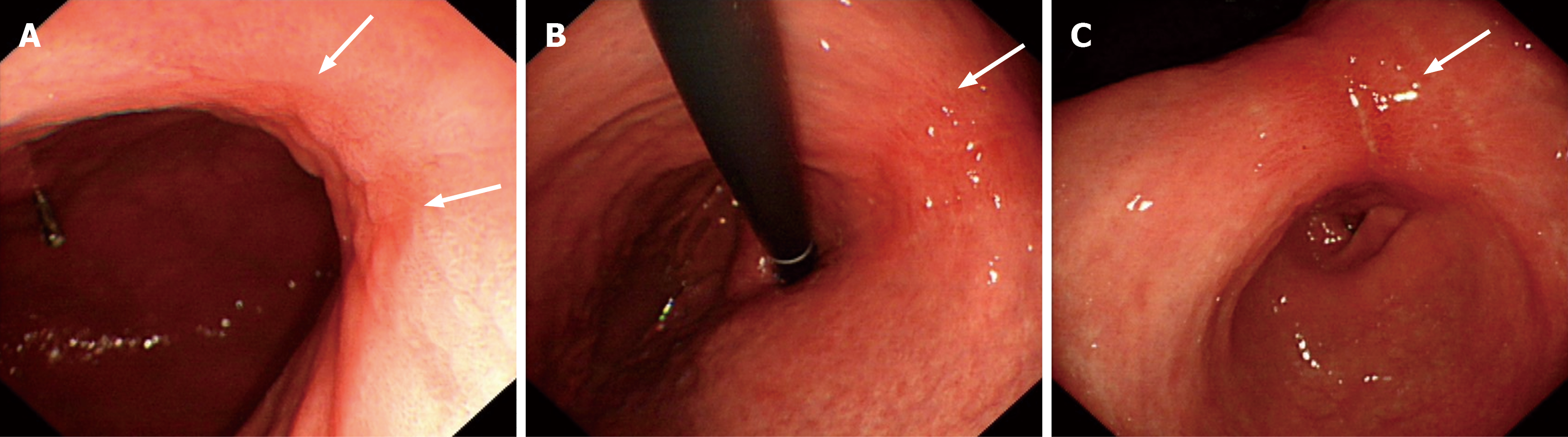
Figure 3 Endoscopy images of the reexamination 4 months after endoscopic submucosal dissection.
A-C: An endoscopic submucosal dissection scar in the gastric body and gastric angulus was observed by the gastroscopy four months after the operation, indicating a well-recovered resection defect without postoperative bleeding (arrows).
- Citation: Chen YM, Xu ZL, Wang LS, Zou ZY. Sprayed tissue adhesive prevents post-endoscopic submucosal dissection bleeding in massive early gastric cancer: A case report. World J Gastrointest Endosc 2026; 18(9): 124149
- URL: https://www.wjgnet.com/1948-5190/full/v18/i9/124149.htm
- DOI: https://dx.doi.org/10.4253/wjge.124149