
















Copyright: ©Author(s) 2026.
World J Gastrointest Endosc. Nov 16, 2026; 18(11): 125857
Published online Nov 16, 2026. doi: 10.4253/wjge.125857
Published online Nov 16, 2026. doi: 10.4253/wjge.125857
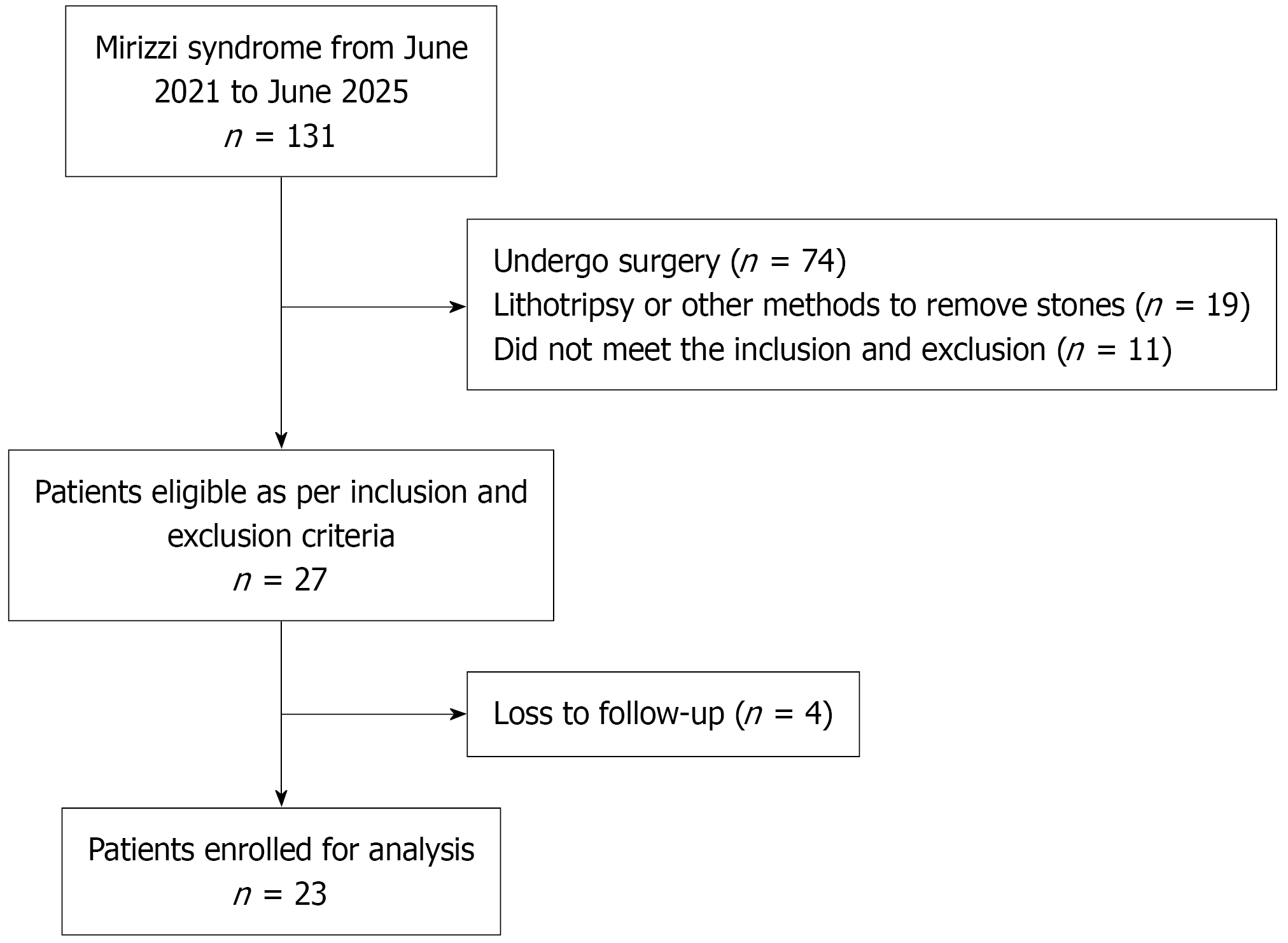
Figure 1 Study flow chart.
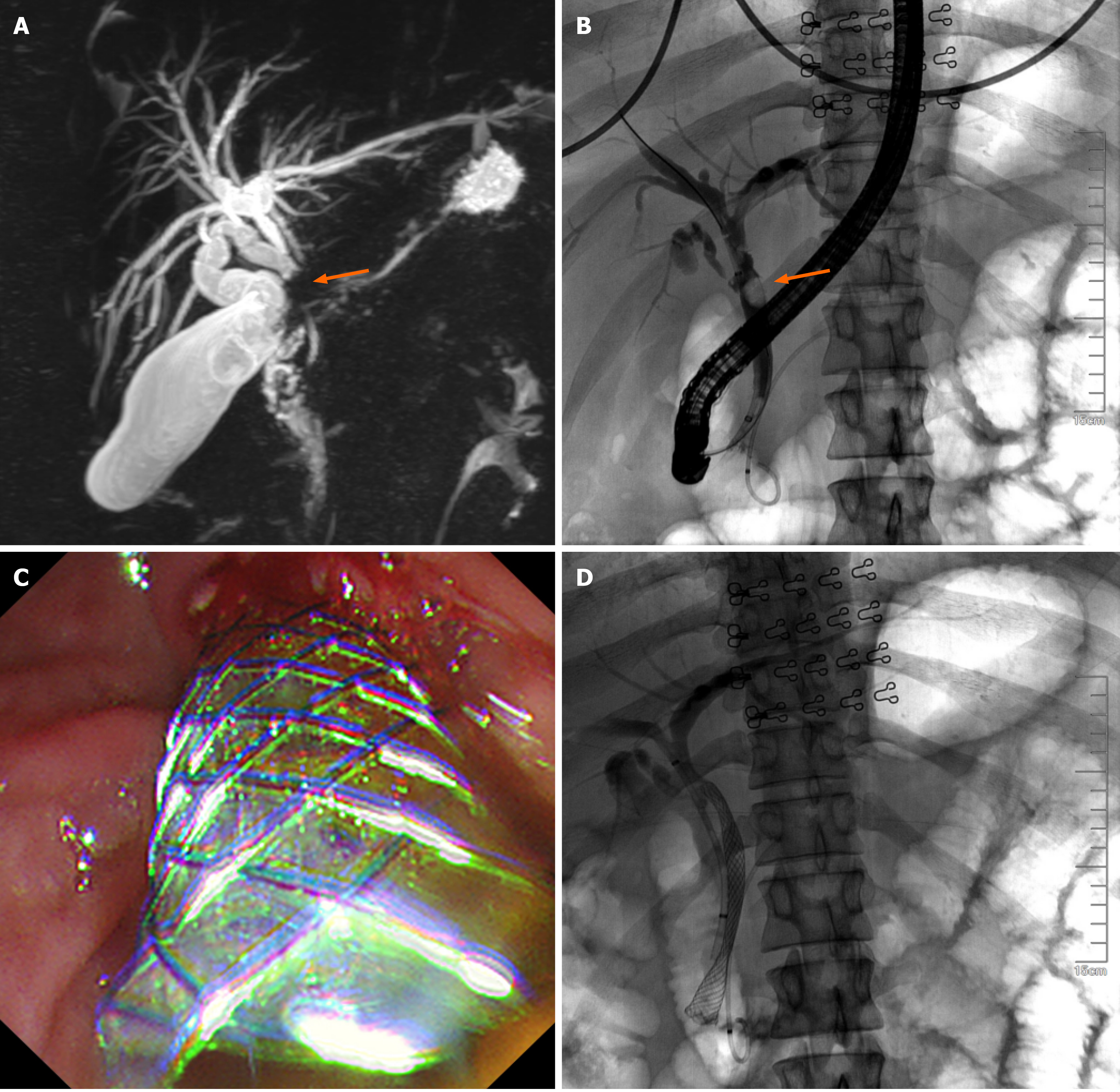
Figure 2 Fully covered biliary stents were placed after evaluation by magnetic resonance cholangiopancreatography and cho
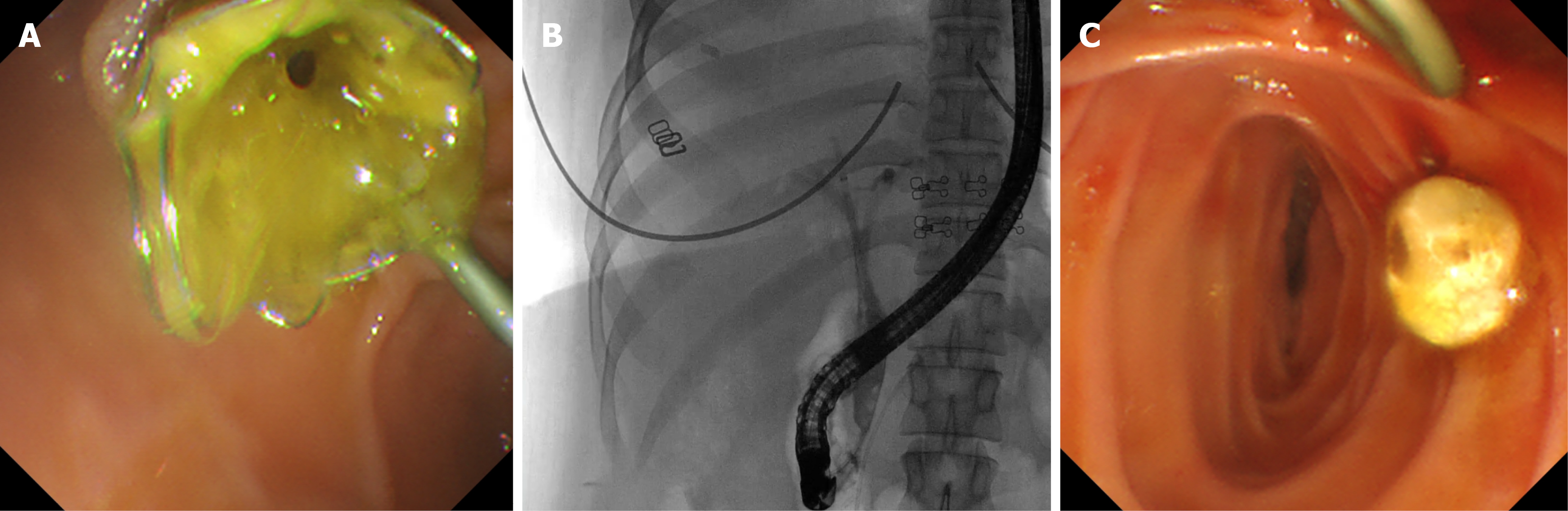
Figure 3 Fully covered stent removal and stone extraction (step 2).
A: Fully covered stent after placement for 8 weeks; B: Cholangiography after fully covered stent removal; C: Removed common bile duct stone.
- Citation: Zou L, Li XC, Yang Y, Huang S, Qin LC. Two-step approach: Stone extraction after covered stent placement in Mirizzi syndrome: Early experience from a retrospective study. World J Gastrointest Endosc 2026; 18(11): 125857
- URL: https://www.wjgnet.com/1948-5190/full/v18/i11/125857.htm
- DOI: https://dx.doi.org/10.4253/wjge.125857