
















Copyright: ©Author(s) 2026.
World J Gastrointest Endosc. Nov 16, 2026; 18(11): 125797
Published online Nov 16, 2026. doi: 10.4253/wjge.125797
Published online Nov 16, 2026. doi: 10.4253/wjge.125797
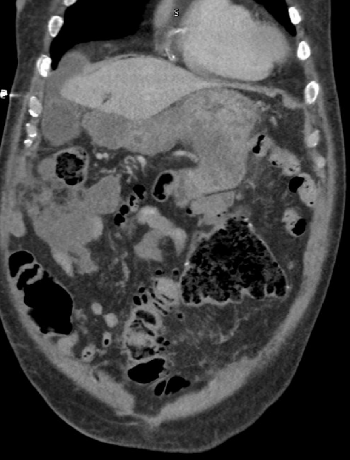
Figure 1
Computed tomography of the abdomen and pelvis showing a large intra-abdominal mass involving the lumen of the duo
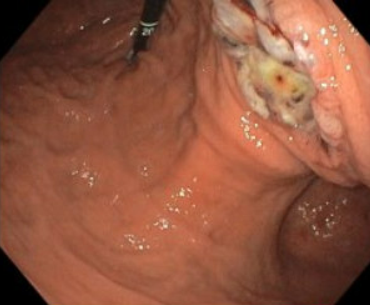
Figure 2
White-light view of the large cratered gastric ulcer with stigmata of recent bleeding in the gastric body at the junction of the greater curvature and posterior wall.
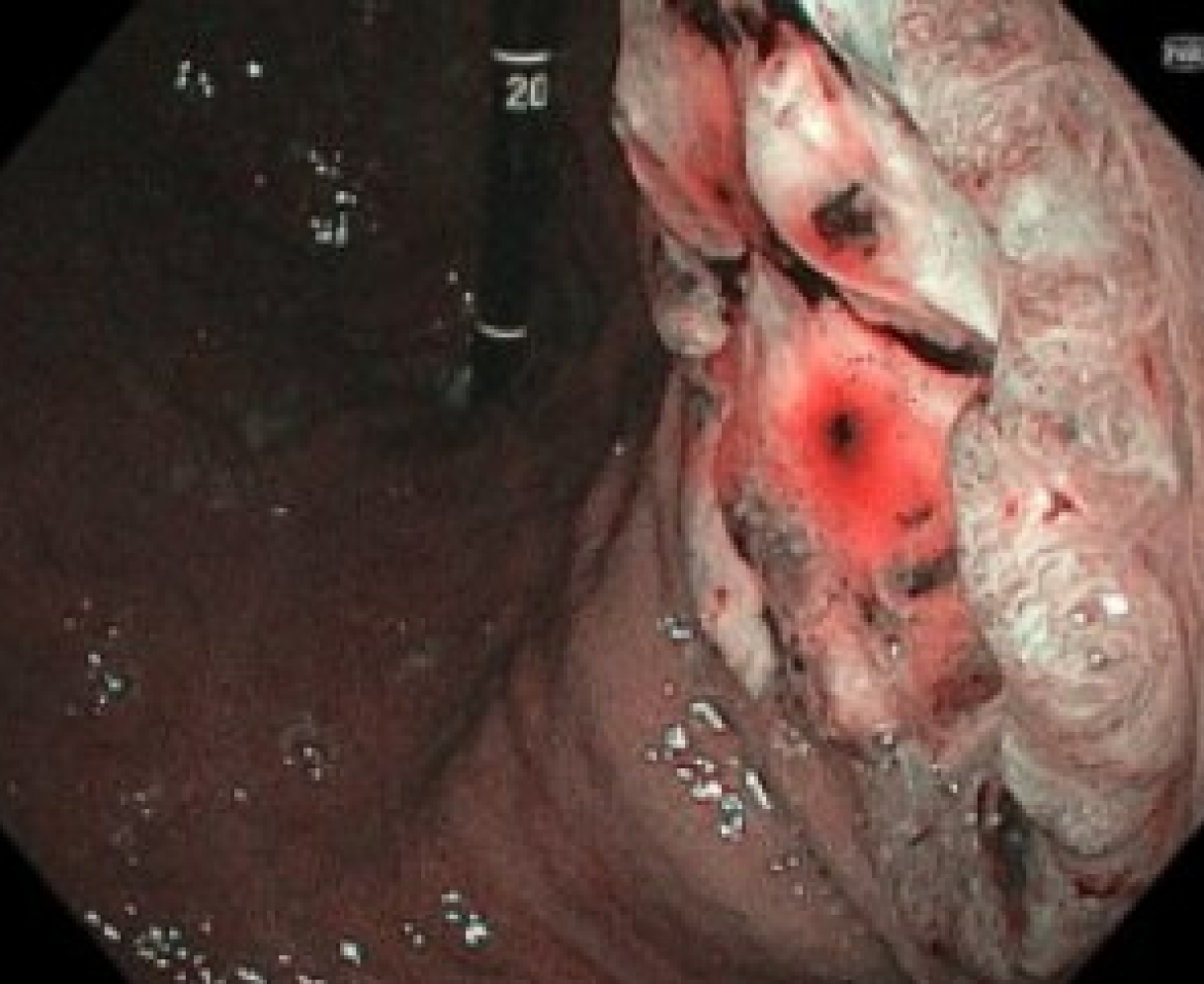
Figure 3
Narrow-band imaging view of the cratered gastric ulcer.
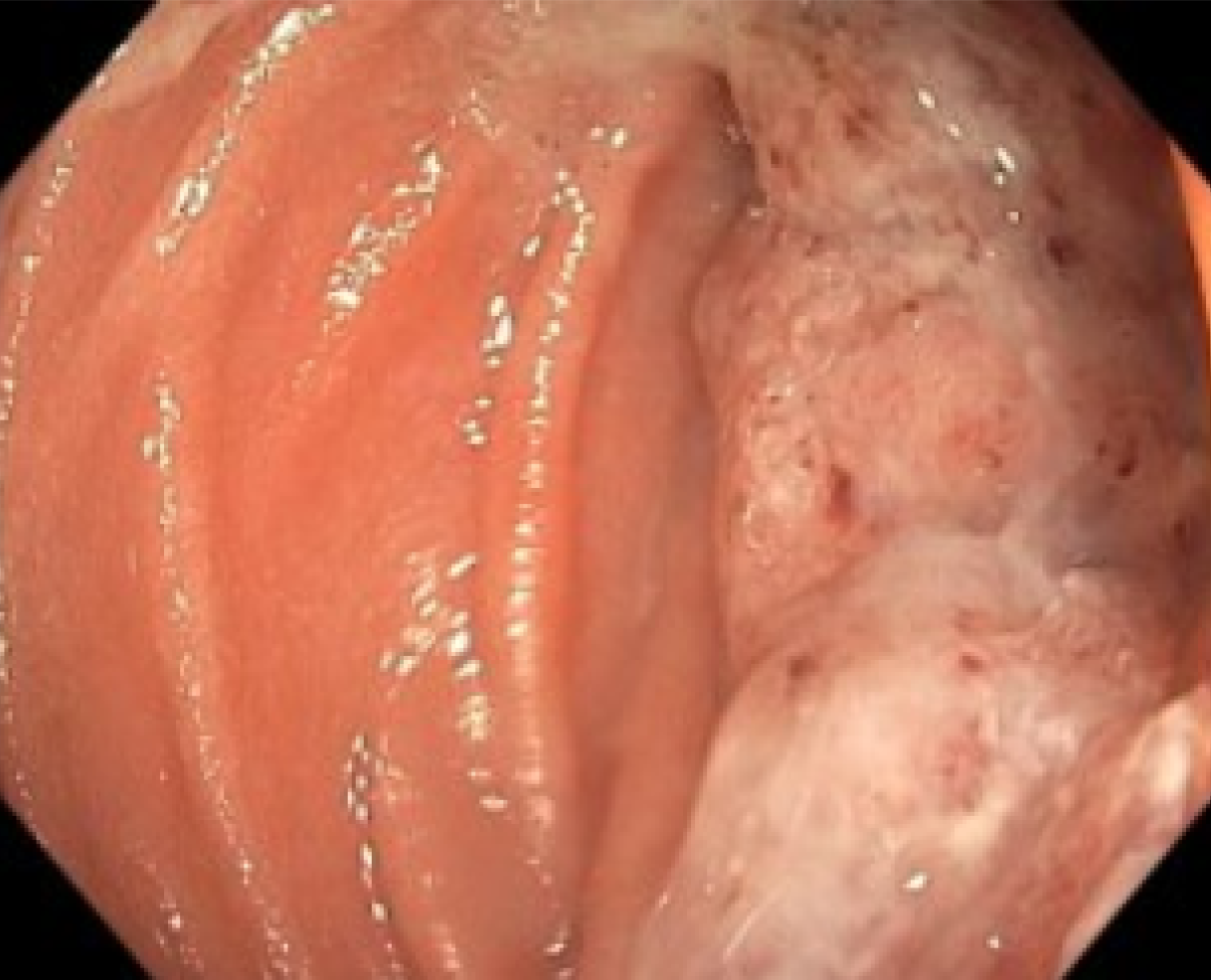
Figure 4
Obstructing mass with overlying mucus in the proximal jejunum.
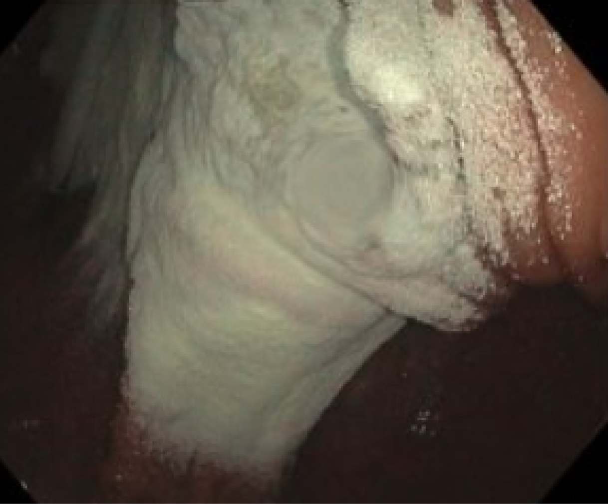
Figure 5
Application of TC-325 to the cratered gastric ulcer with the endoscope in partial retroflexion.
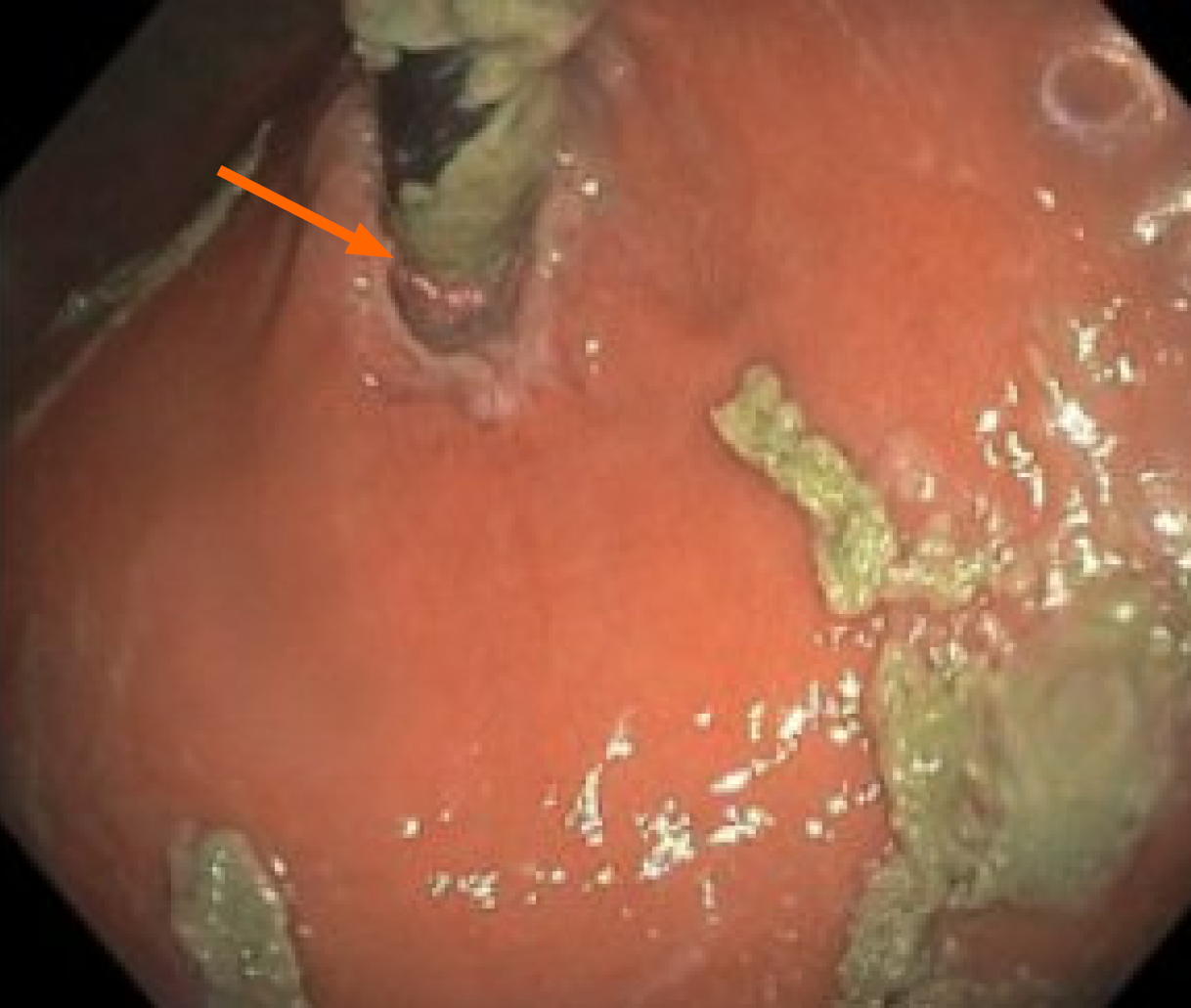
Figure 6 Retroflexed view showing the insertion tube coated with hydrated TC-325 and bonded to the mucosa at the cardia/gas
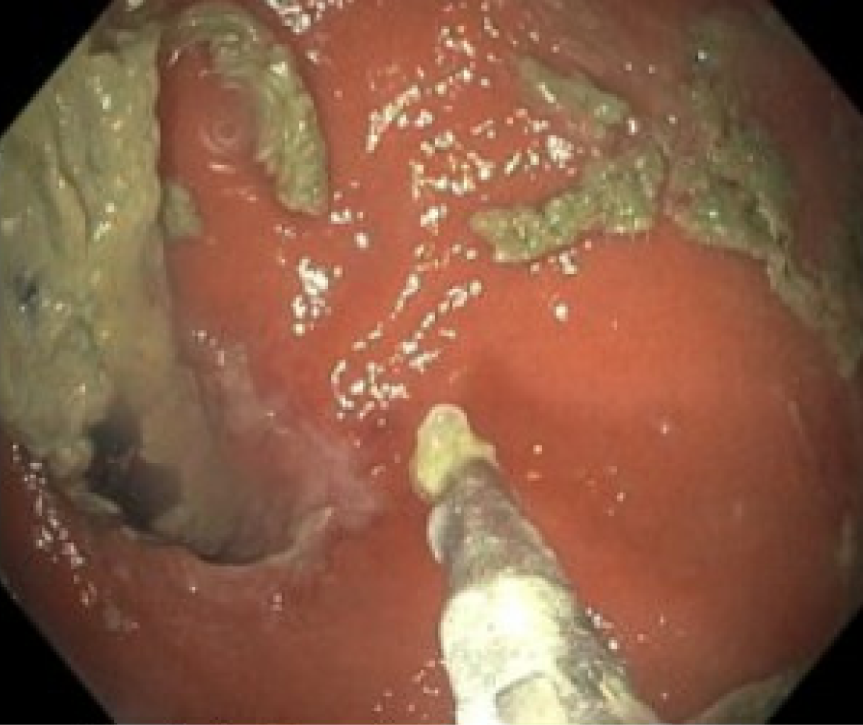
Figure 7
Attempt to remove adherent TC-325 from the insertion tube with biopsy forceps passed through the entrapped endoscope in the retroflexed view.
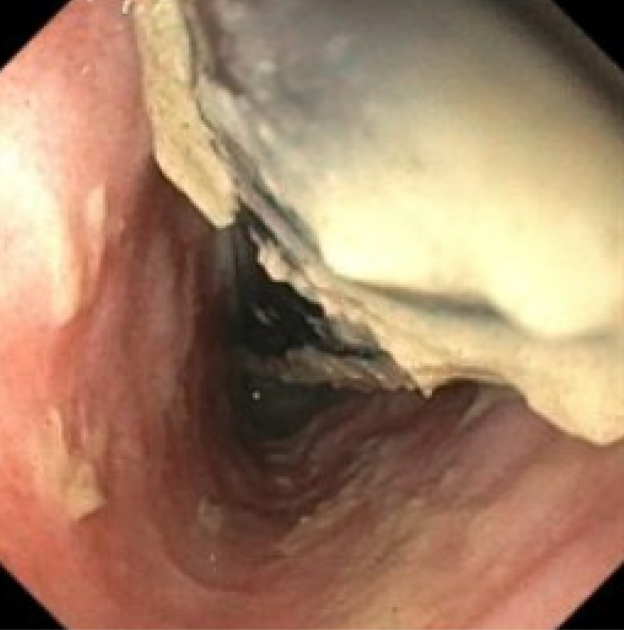
Figure 8
Ultrathin (XP) gastroscope advanced alongside the entrapped pediatric colonoscope; biopsy forceps passed through the ultrathin scope are used to clear the adherent TC-325 from the insertion tube under direct vision.
- Citation: Gangu K, Daglilar E, Chela HK. Stuck scope: Tackling the adherent scope situation after hemostatic powder application: A case report and review of literature. World J Gastrointest Endosc 2026; 18(11): 125797
- URL: https://www.wjgnet.com/1948-5190/full/v18/i11/125797.htm
- DOI: https://dx.doi.org/10.4253/wjge.125797