Copyright
©The Author(s) 2019.
World J Gastrointest Endosc. Jun 16, 2019; 11(6): 403-412
Published online Jun 16, 2019. doi: 10.4253/wjge.v11.i6.403
Published online Jun 16, 2019. doi: 10.4253/wjge.v11.i6.403
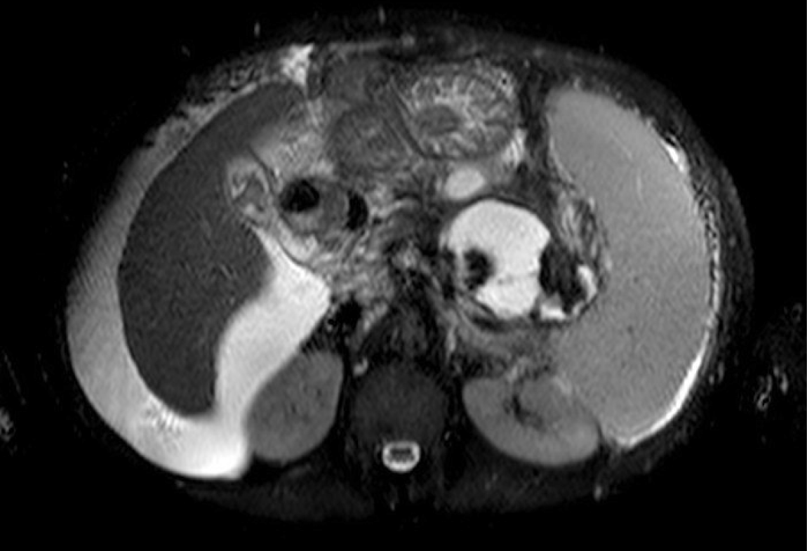
Figure 1 An axial T2 weighted magnetic resonance image showing walled-off necrosis, cirrhotic liver and ascites.
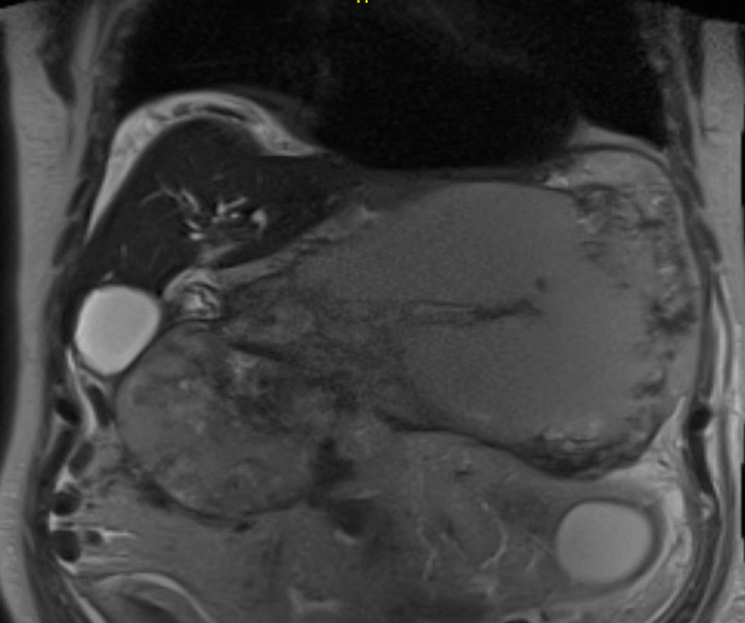
Figure 2 A coronal magnetic resonance image showing a large walled-off necrosis, cirrhotic liver and ascites.
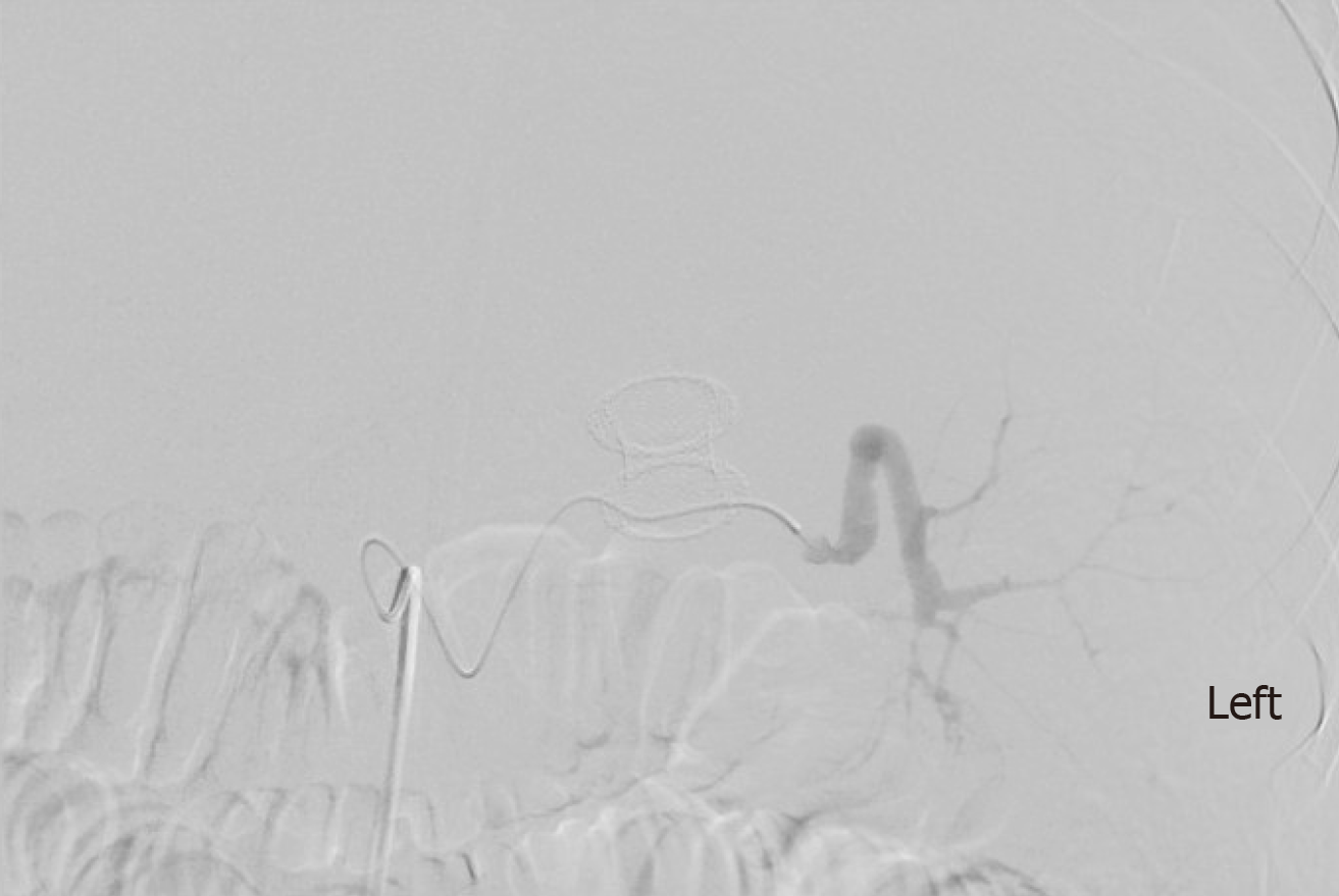
Figure 3 Angiogram showing ruptured splenic artery pseudoaneurysm and lumen-apposing self-expandable metal stents.
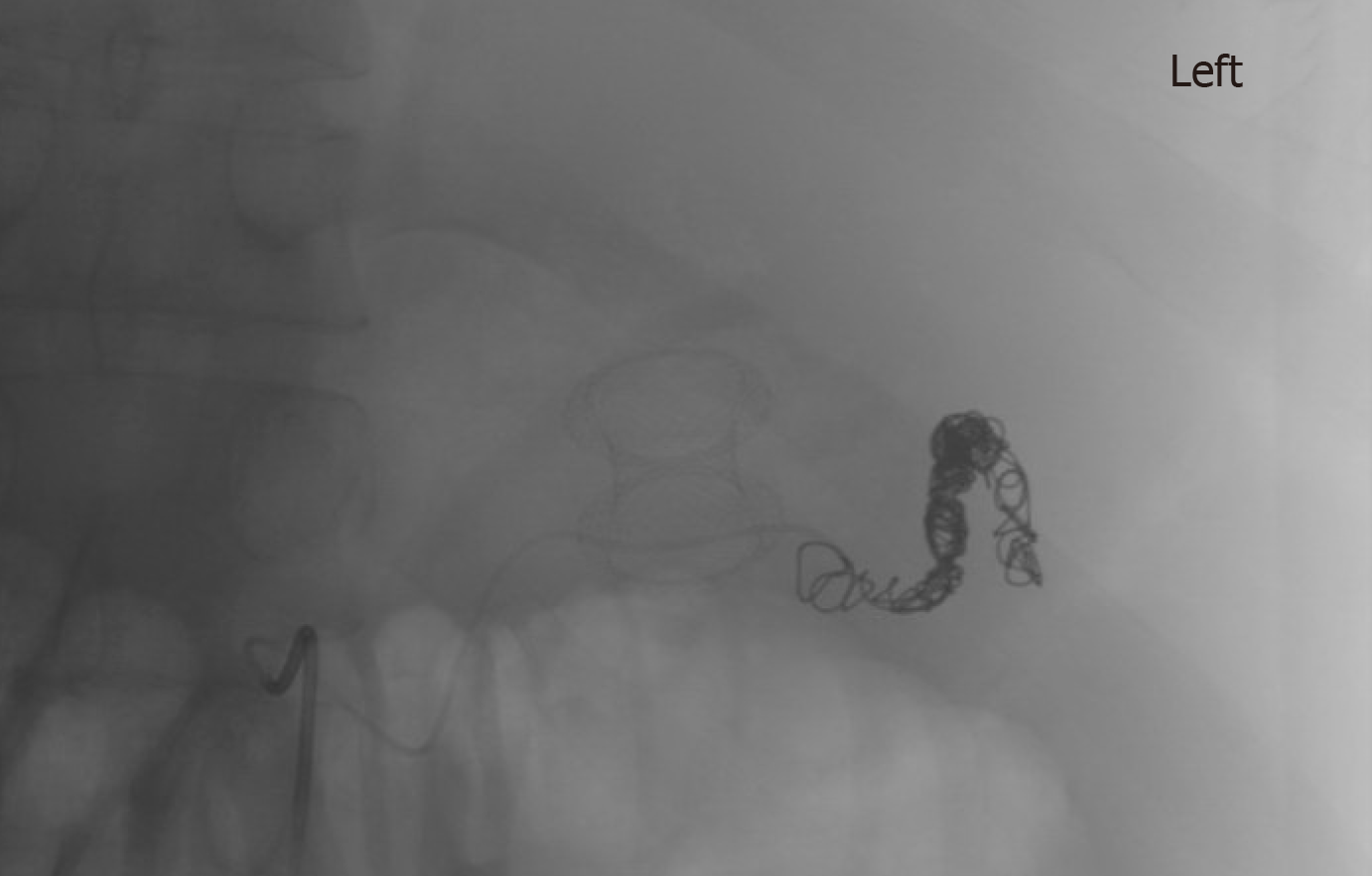
Figure 4 Selective embolization of splenic artery pseudoaneurysm and lumen-apposing self-expandable metal stents.
- Citation: Laique S, Franco MC, Stevens T, Bhatt A, Vargo JJ, Chahal P. Clinical outcomes of endoscopic management of pancreatic fluid collections in cirrhotics vs non-cirrhotics: A comparative study. World J Gastrointest Endosc 2019; 11(6): 403-412
- URL: https://www.wjgnet.com/1948-5190/full/v11/i6/403.htm
- DOI: https://dx.doi.org/10.4253/wjge.v11.i6.403


