Copyright
©The Author(s) 2019.
World J Gastrointest Endosc. Apr 16, 2019; 11(4): 308-321
Published online Apr 16, 2019. doi: 10.4253/wjge.v11.i4.308
Published online Apr 16, 2019. doi: 10.4253/wjge.v11.i4.308
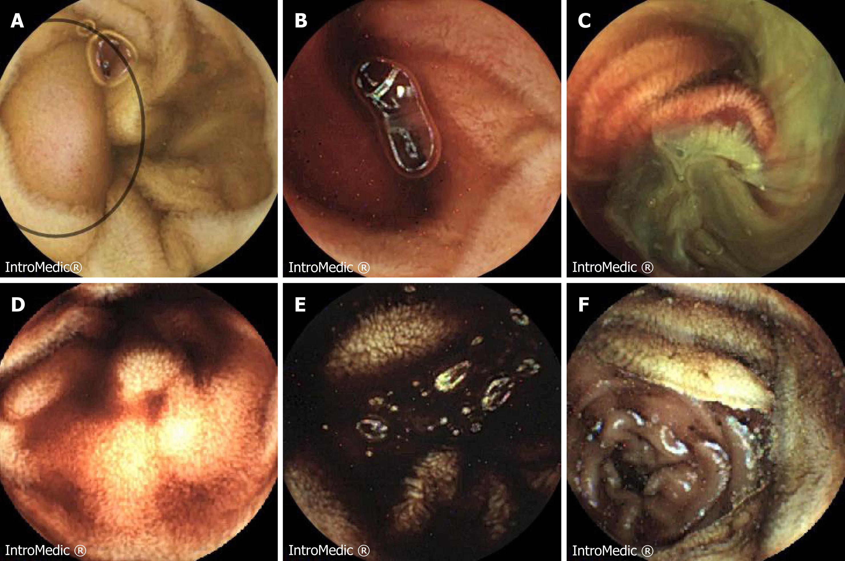
Figure 1 Images from small bowel capsule endoscopy.
A: Images from small bowel capsule endoscopy demonstrates a submucosal lesion in the mid-small bowel; B-D: It demonstrates in close proximity to active bleeding, which was likely to be the cause of the patient’s presenting complain; E, F: Progressively darker bleeding towards the rest of the ileum turning to melena.
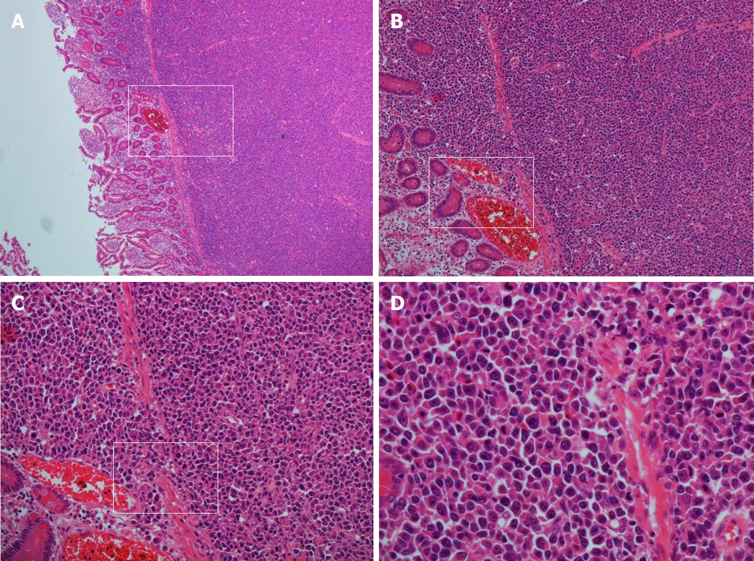
Figure 2 Haematoxylin and eosin staining.
Infiltration with sheets of neoplastic pleomorphic cells with plasmacytoid appearance involving the full thickness of the bowel wall. Plasmablasts have highly atypical nuclei with prominent nucleoli. Magnifications: A: ×4; B: ×10; C: ×20; D: ×40.
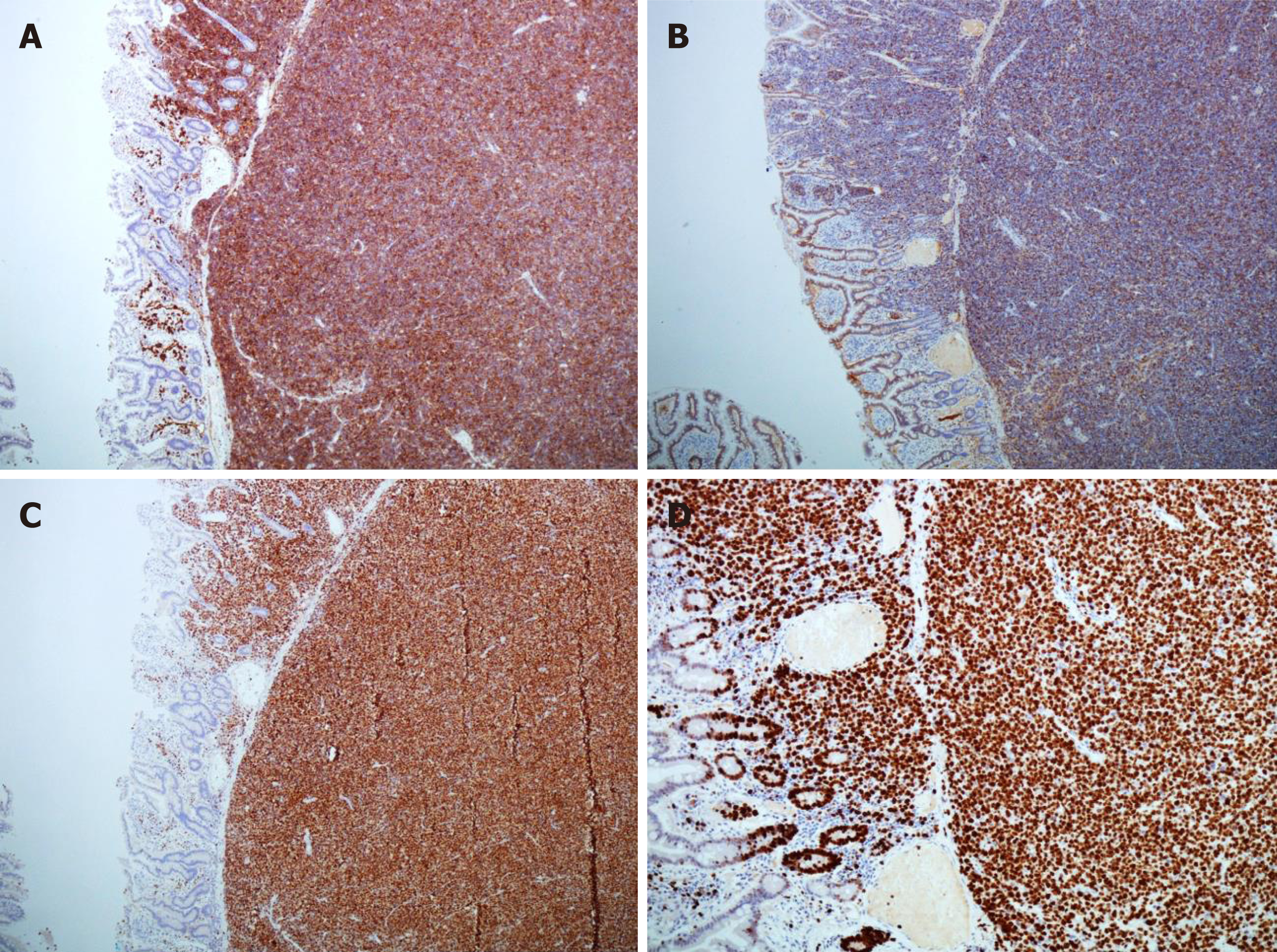
Figure 3 Small bowel plasmablastic myeloma staining.
A: Small bowel plasmablastic myeloma positive CD38 staining. CD38 is routinely used for identification of plasma cell neoplasms and stains primarily the membrane due to expression of the transmembrane protein cyclic ADP ribose hydrolase; B: Same specimen positive for CD 138 staining. CD138 is a transmembrane heparan sulphate proteoglycan (syndecan-1). CD138 staining is positive in normal B-cell precursors and plasma cells, along as plasmablastic lymphomas and myelomas; C: Same specimen positive for MUM-1 (Multiple Myeloma-1) nuclear stain. MUM-1 is a nuclear transcriptional factor that is expressed in late plasma cell directed stages of B cell differentiation and also in activated T cells; D: Small bowel plasmablastic myeloma staining positive for Ki 67 (MIB-1). MIB-1 is the IgG1 antibody against Ki 67 which can be detected in the cellular nucleus and is a marker of cell proliferation. Strong expression of the nuclear marker Ki 67 with MIB-1 staining indicates high proliferation rate and is a sign of clinical aggressiveness.
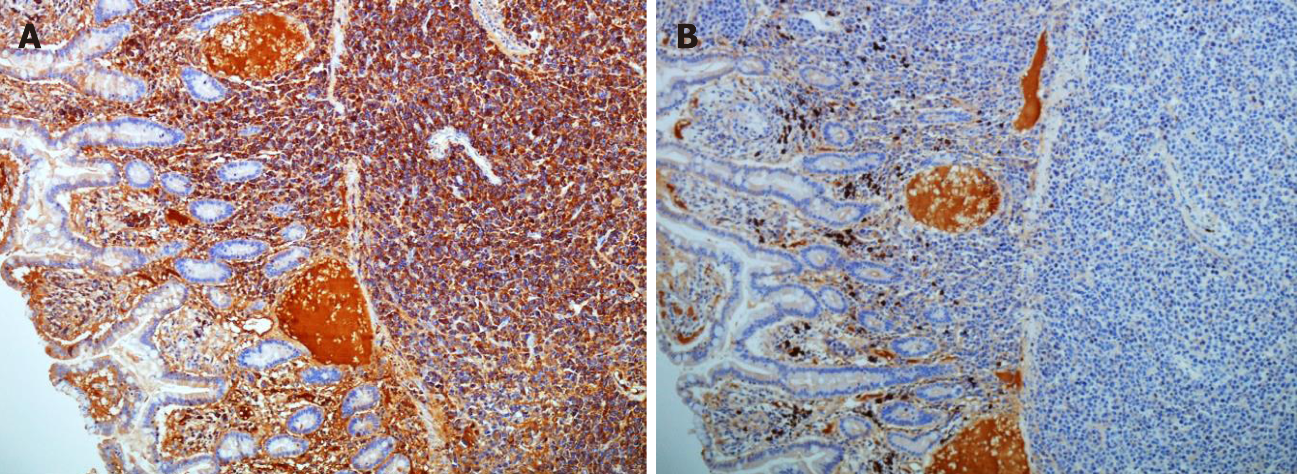
Figure 4 Small bowel plasmablastic myeloma specimen.
A: Small bowel plasmablastic myeloma specimen demonstrating strong staining with lambda in keeping with lambda light chain restriction; B: Same specimen showing very weak staining with kappa.
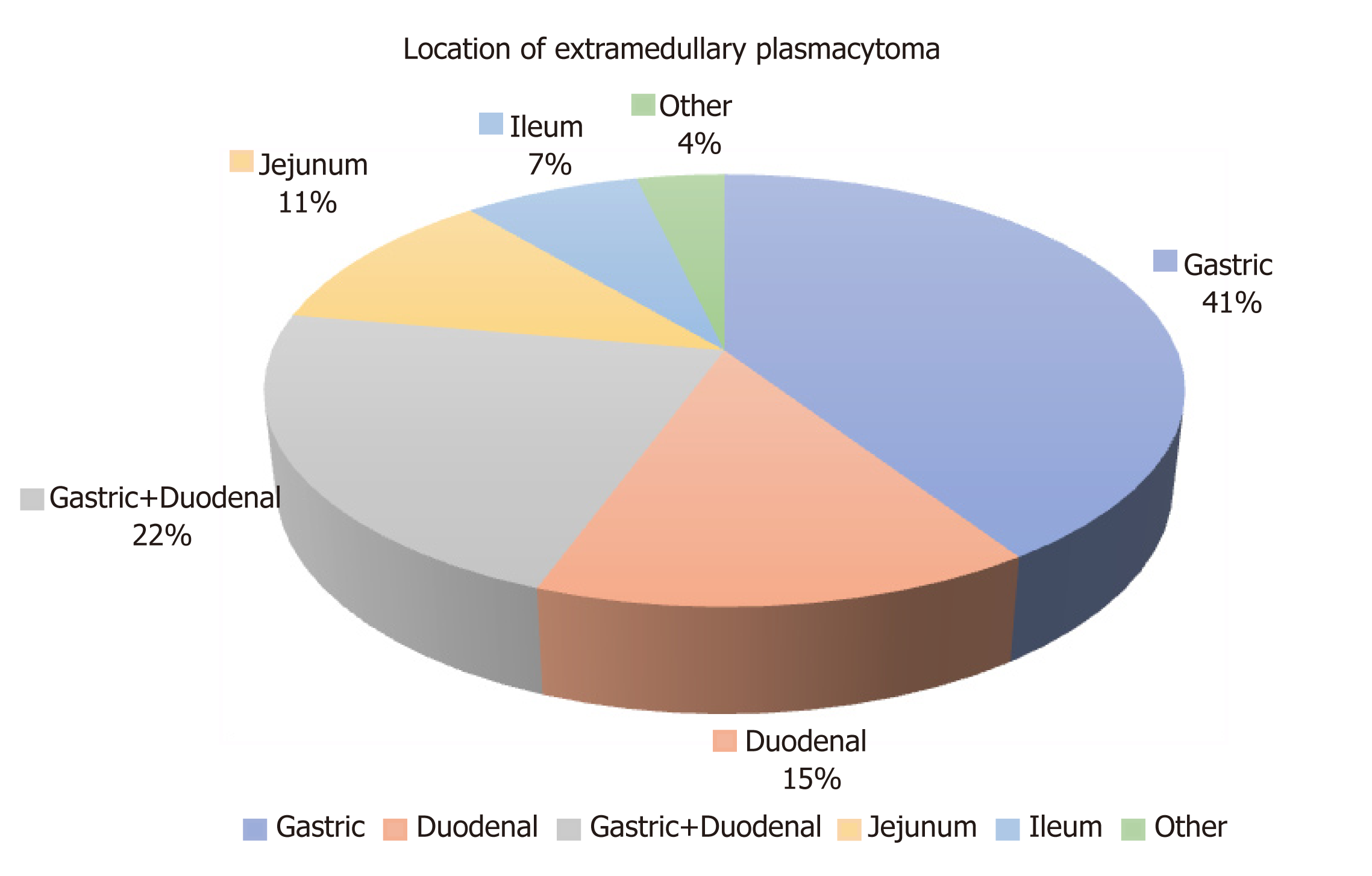
Figure 5 Location in the gastrointestinal tract of the extramedullary plasma cell neoplasms in the case reports reviewed.
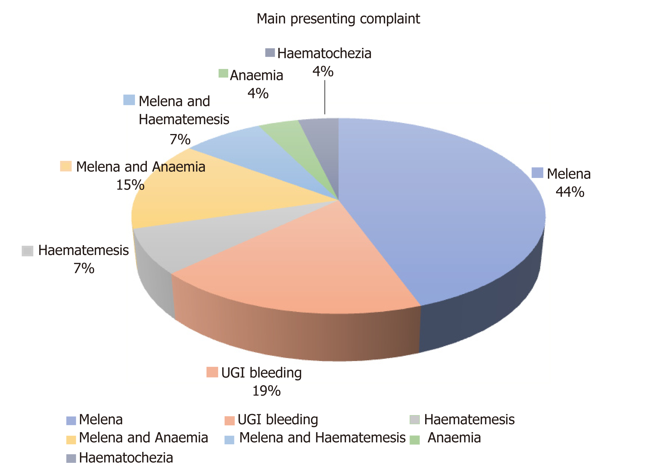
Figure 6 Main presenting complaints described in the case reports reviewed.
UGI: Upper gastrointestinal.
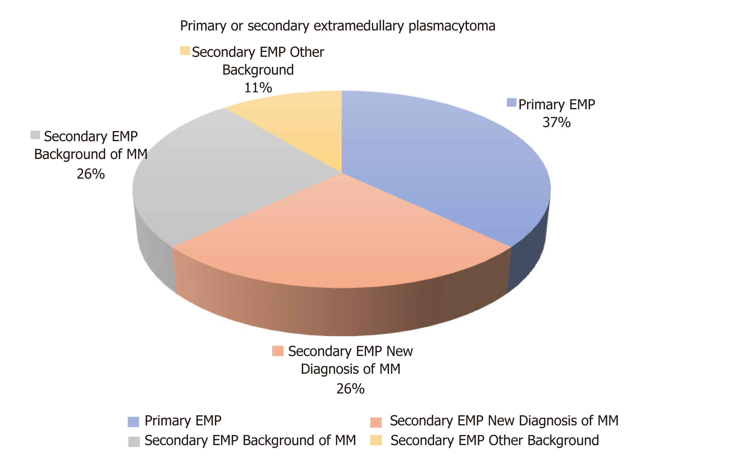
Figure 7 Primary of secondary nature of the extramedullary plasmacytomas described in the case reports reviewed.
EMP: Extramedullary plasmacytoma; MM: Multiple myeloma.
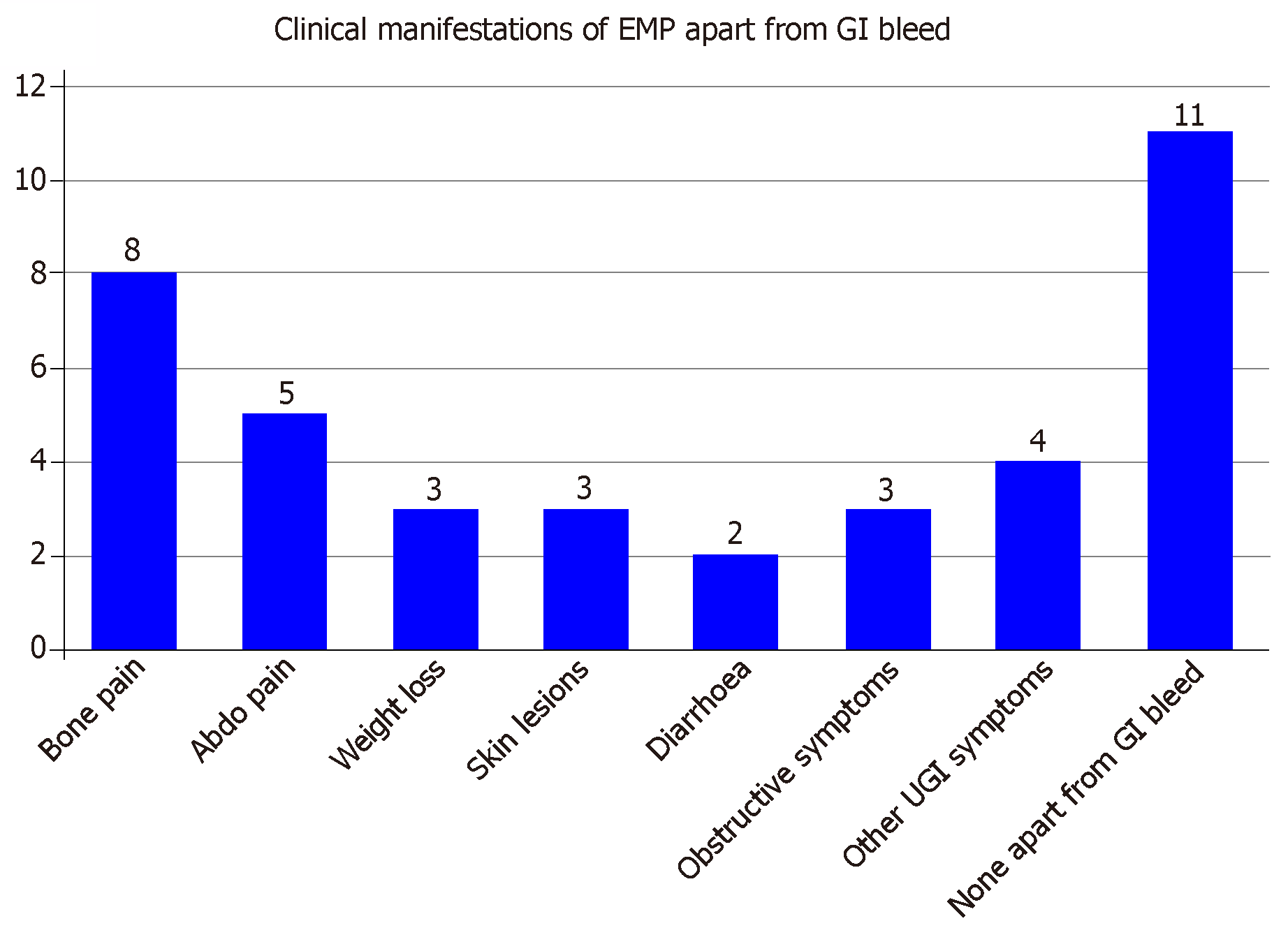
Figure 8 Associated clinical manifestations of extramedullary plasmacytomas apart from gastrointestinal bleeding described in the case reports reviewed.
GI: gastrointestinal; EMP: Extramedullary plasmacytoma; UGI: Upper gastrointestinal.
- Citation: Iosif E, Rees C, Beeslaar S, Shamali A, Lauro R, Kyriakides C. Gastrointestinal bleeding as initial presentation of extramedullary plasma cell neoplasms: A case report and review of the literature. World J Gastrointest Endosc 2019; 11(4): 308-321
- URL: https://www.wjgnet.com/1948-5190/full/v11/i4/308.htm
- DOI: https://dx.doi.org/10.4253/wjge.v11.i4.308


