Copyright
©The Author(s) 2019.
World J Gastrointest Endosc. Apr 16, 2019; 11(4): 298-307
Published online Apr 16, 2019. doi: 10.4253/wjge.v11.i4.298
Published online Apr 16, 2019. doi: 10.4253/wjge.v11.i4.298
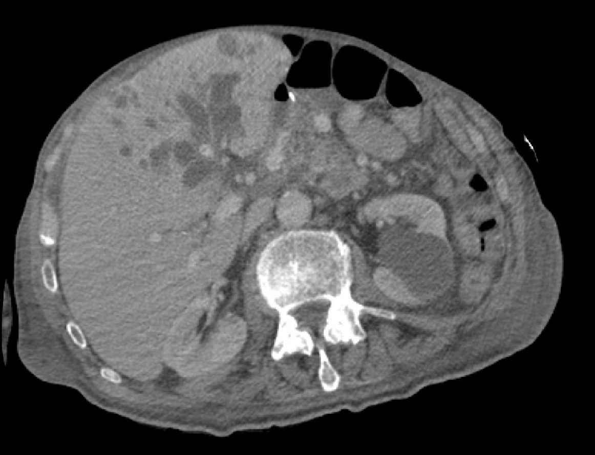
Figure 1 Computerised tomography scan image in Patient 1 showing dilated segment 4, 2 and 3 ducts.
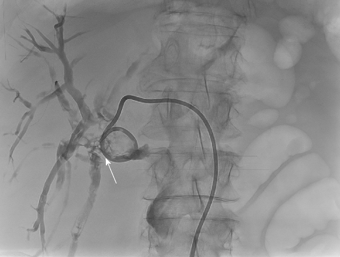
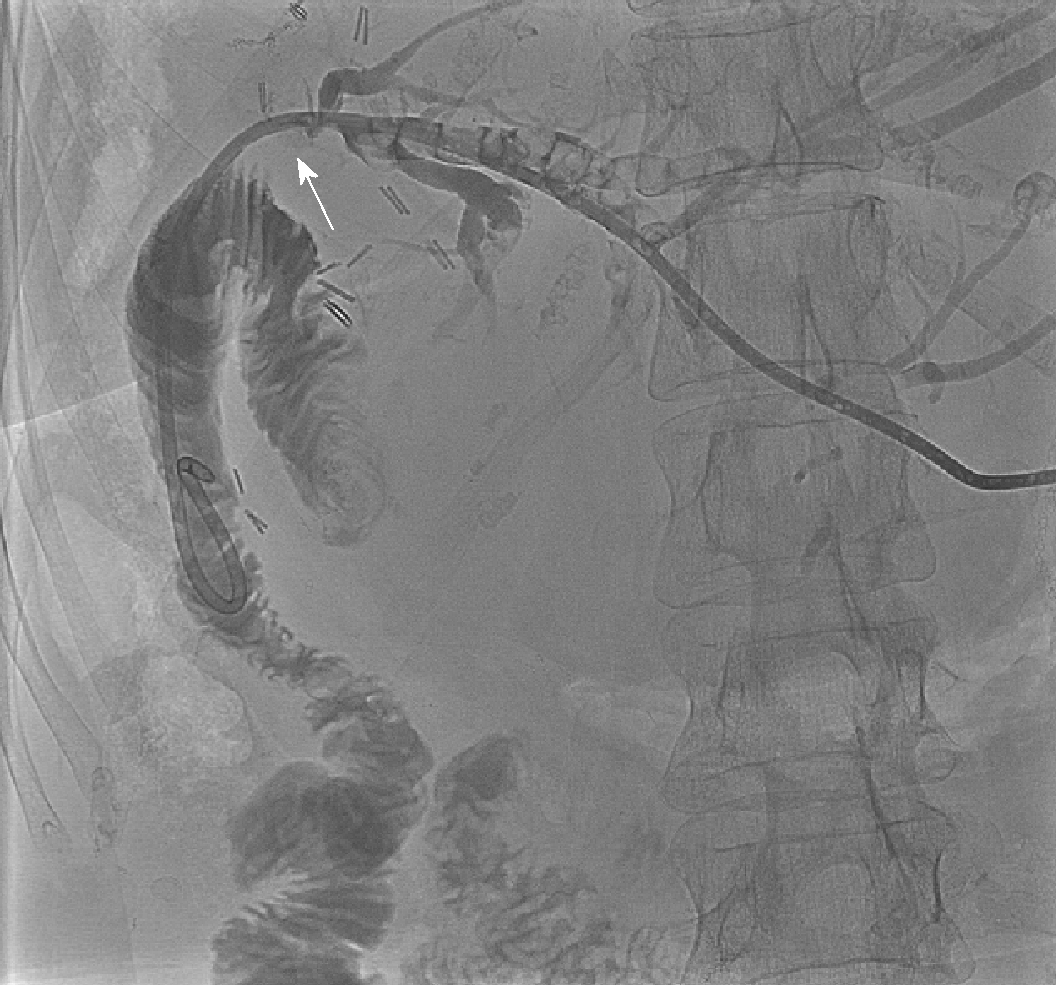
Figure 2 Percutaneous transhepatic cholangiography via segment 3 duct in Patient 1 showing filling defect consistent with an obstructing extrahepatic calculus (white arrow) at origin of the left hepatic ducts and located just proximal to the choledochoduodenostomy anastomosis.
An external biliary drain is inserted.
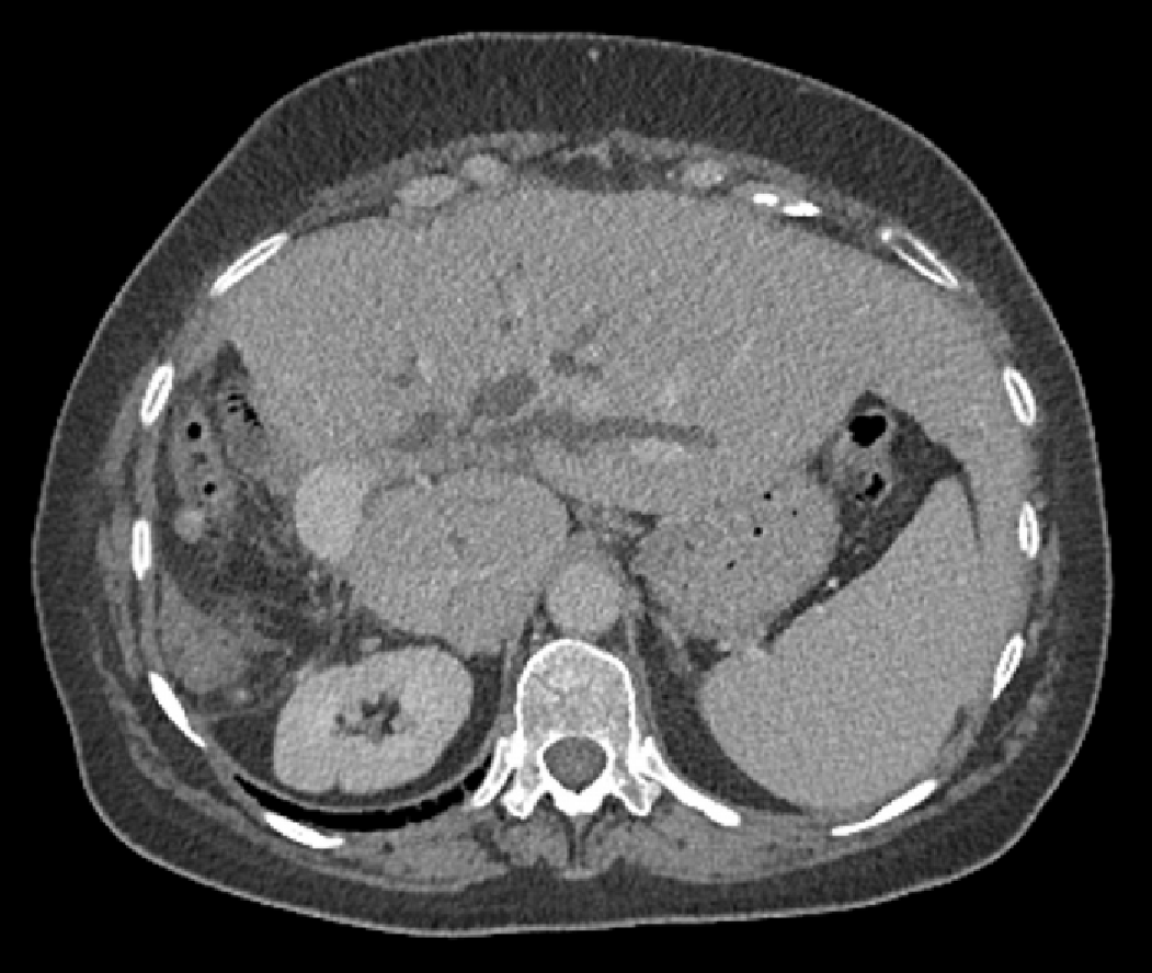
Figure 3 Computerised tomography scan image in Patient 2 showing previous right hemihepatectomy and dilated left intrahepatic ducts.
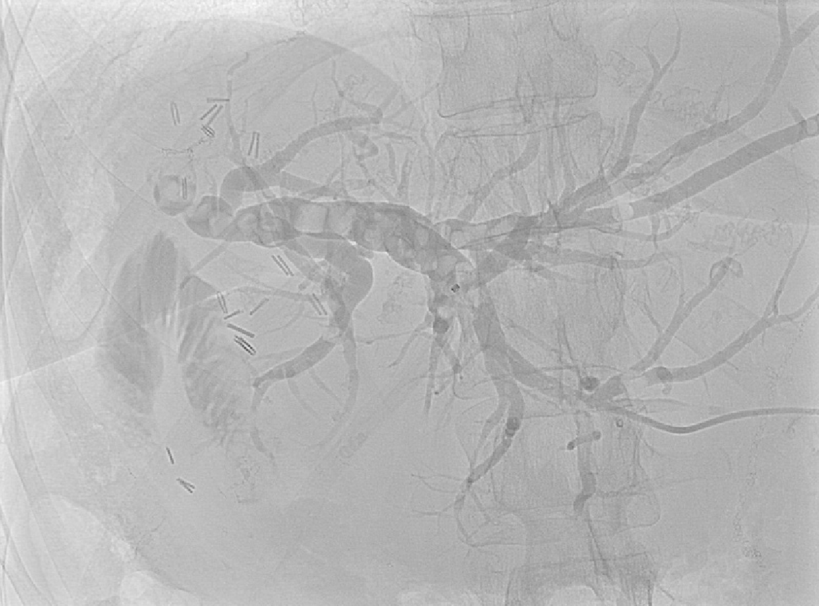
Figure 4 Percutaneous transhepatic cholangiography shows dilated left intrahepatic in addition to peripheral segment 3 ducts containing multiple calculi.
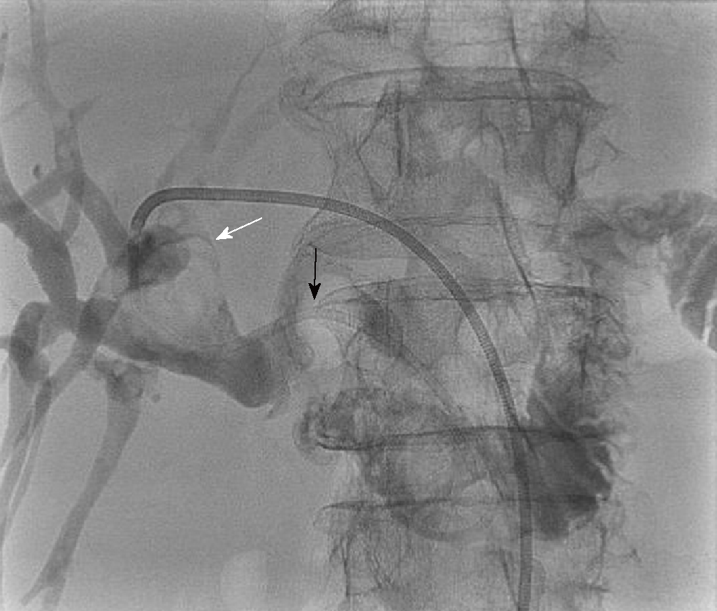
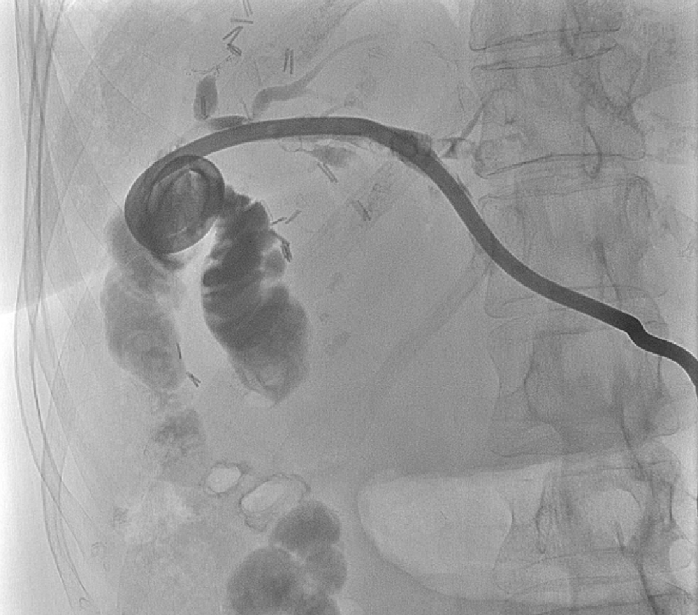
Figure 5 The external percutaneous transhepatic cholangiography drain was converted to an internal-external drain (black arrow) 6 d following the original percutaneous transhepatic cholangiography.
The obstructing stone (white arrow) is again seen in the same position as in Figure 2. The distal locking loop of the drain is left in the duodenum and thus permits entry of contrast into the small bowel.
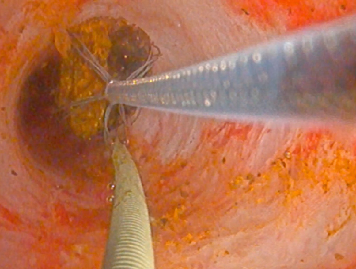
Figure 6 Video cholangioscopy image showing a guide wire (on left side of image) in the bile duct and a mobile stone being basket-retrieved from bile duct using an introduced Dorma basket (on right side of image).
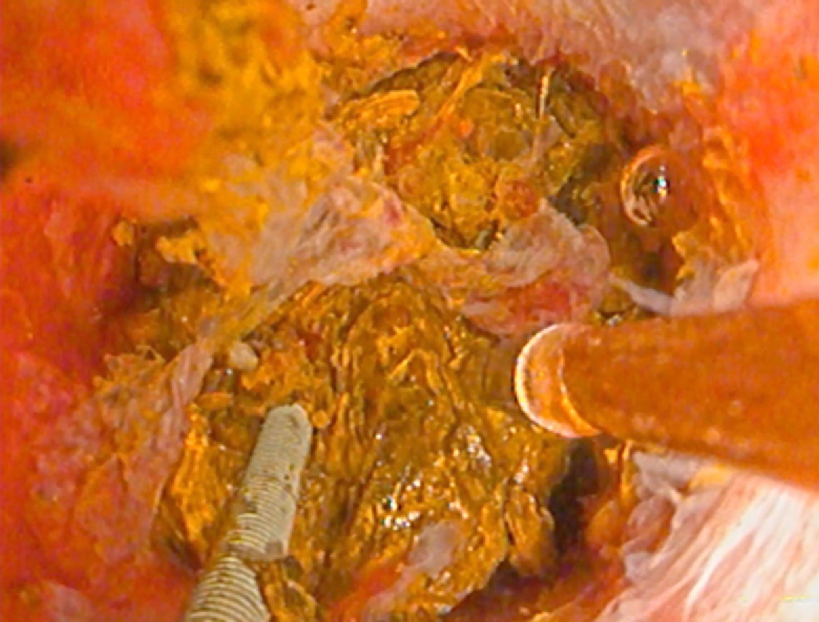
Figure 7 Video cholangioscopy image showing guide wire (on left side of image) in bile duct lumen alongside impacted stone cluster with lithotripsy probe introduced (on right side of image) for electrohydraulic stone fragmentation.
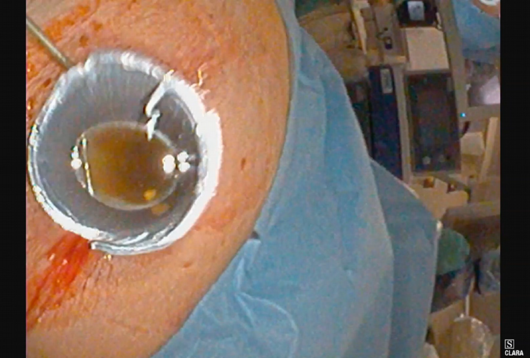
Figure 8 Percutaneous transhepatic cholangioscopy and lithotripsy for ductal calculi.
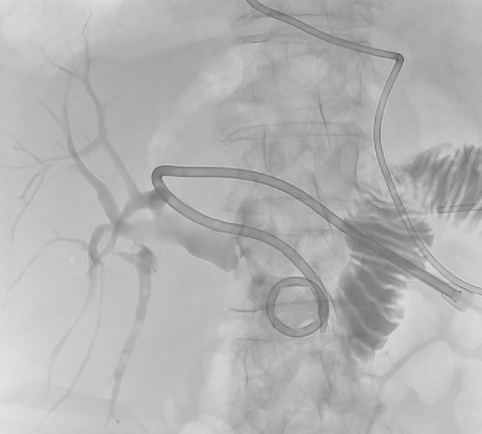
Figure 9 Percutaneous transhepatic cholangiography image for Patient 2 demonstrating the internal-external biliary drain (arrowed) traversing the hepaticojejunostomy stricture so contrast is now seen in the jejunum.
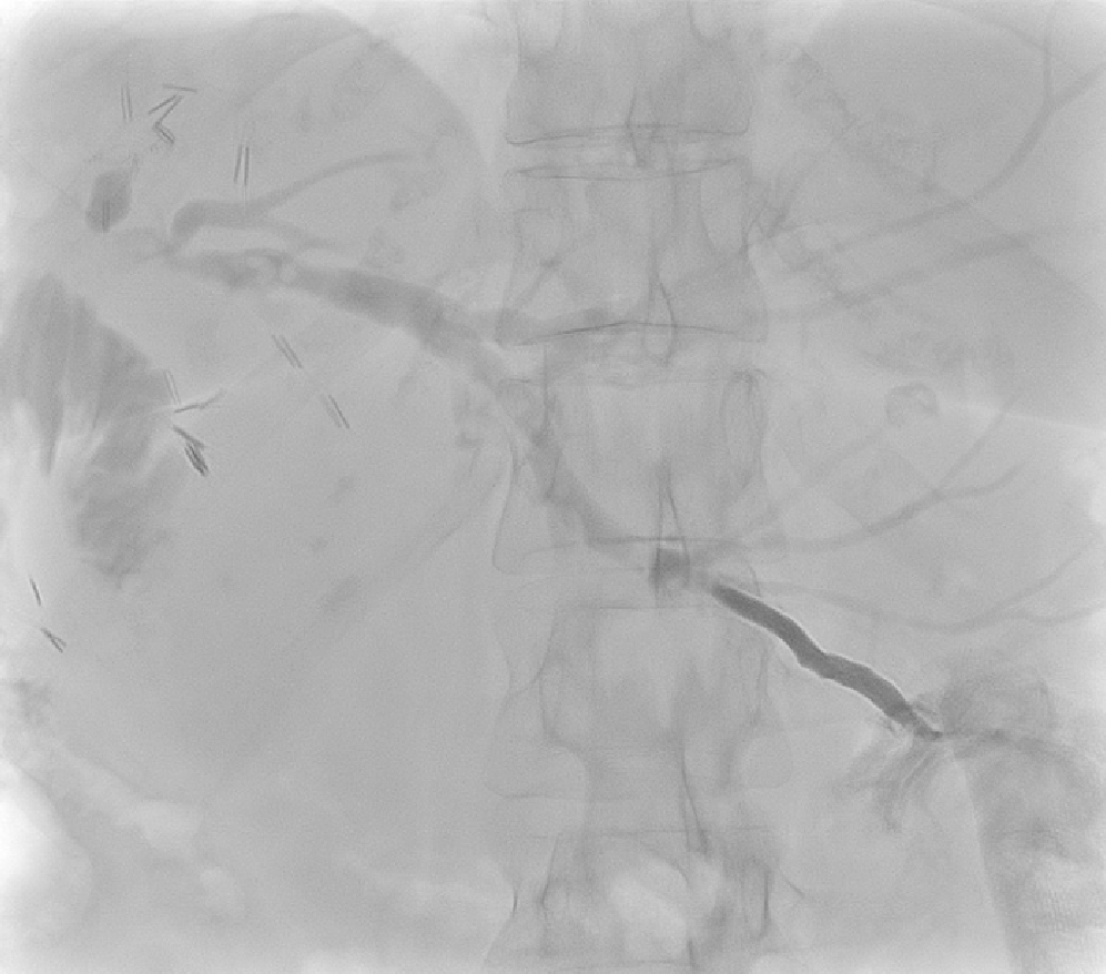
Figure 10 Post-percutaneous transhepatic cholangioscopy and lithotripsy cholangiogram in Patient 1 showing unobstructed bile ducts but few small calculi in Segment 6.
Internalised percutaneous transhepatic cholangiography drain is seen with distal locked end in the duodenum.
Figure 11 Post-percutaneous transhepatic cholangioscopy and lithotripsy check cholangiogram after in Patient 2 showed stones in segment 2.
Figure 12 Cholangiogram performed after repeat procedure in Patient 2 to flush remnant stones showed smaller and fewer remnant intrahepatic biliary duct stones.
- Citation: Alabraba E, Travis S, Beckingham I. Percutaneous transhepatic cholangioscopy and lithotripsy in treating difficult biliary ductal stones: Two case reports. World J Gastrointest Endosc 2019; 11(4): 298-307
- URL: https://www.wjgnet.com/1948-5190/full/v11/i4/298.htm
- DOI: https://dx.doi.org/10.4253/wjge.v11.i4.298


