
















©The Author(s) 2019.
World J Gastrointest Endosc. Apr 16, 2019; 11(4): 298-307
Published online Apr 16, 2019. doi: 10.4253/wjge.v11.i4.298
Published online Apr 16, 2019. doi: 10.4253/wjge.v11.i4.298
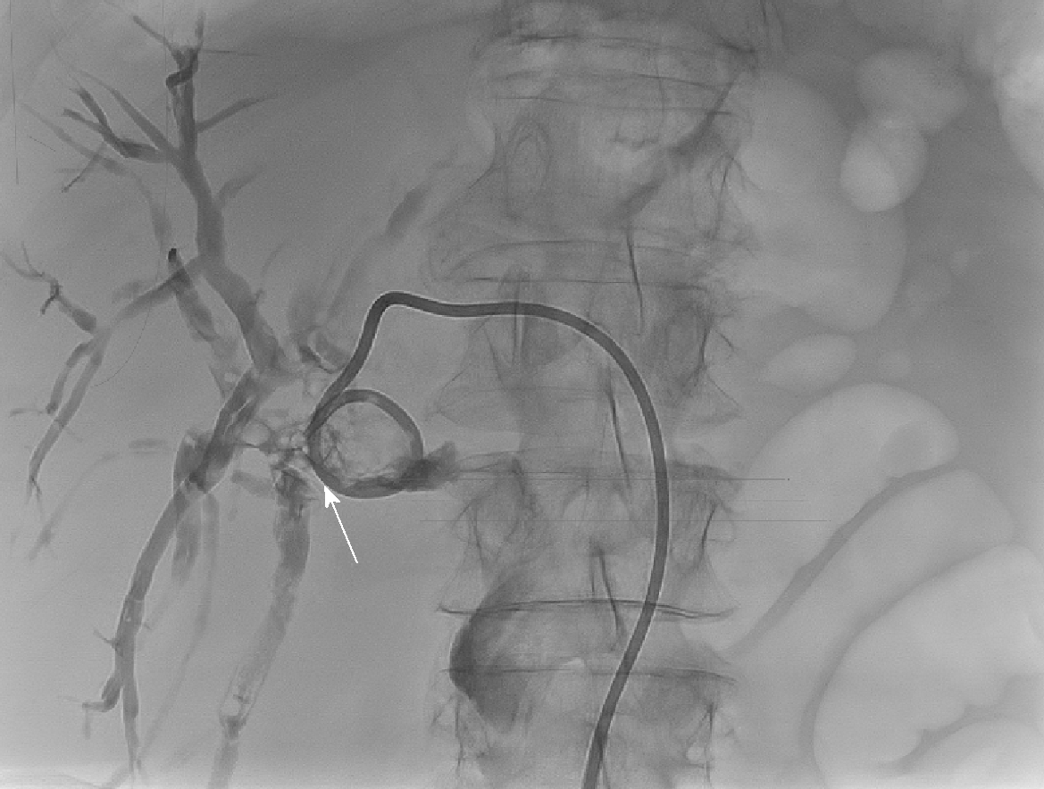
Figure 2 Percutaneous transhepatic cholangiography via segment 3 duct in Patient 1 showing filling defect consistent with an obstructing extrahepatic calculus (white arrow) at origin of the left hepatic ducts and located just proximal to the choledochoduodenostomy anastomosis.
An external biliary drain is inserted.
- Citation: Alabraba E, Travis S, Beckingham I. Percutaneous transhepatic cholangioscopy and lithotripsy in treating difficult biliary ductal stones: Two case reports. World J Gastrointest Endosc 2019; 11(4): 298-307
- URL: https://www.wjgnet.com/1948-5190/full/v11/i4/298.htm
- DOI: https://dx.doi.org/10.4253/wjge.v11.i4.298