Copyright
©The Author(s) 2016.
World J Hepatol. Feb 18, 2016; 8(5): 282-290
Published online Feb 18, 2016. doi: 10.4254/wjh.v8.i5.282
Published online Feb 18, 2016. doi: 10.4254/wjh.v8.i5.282
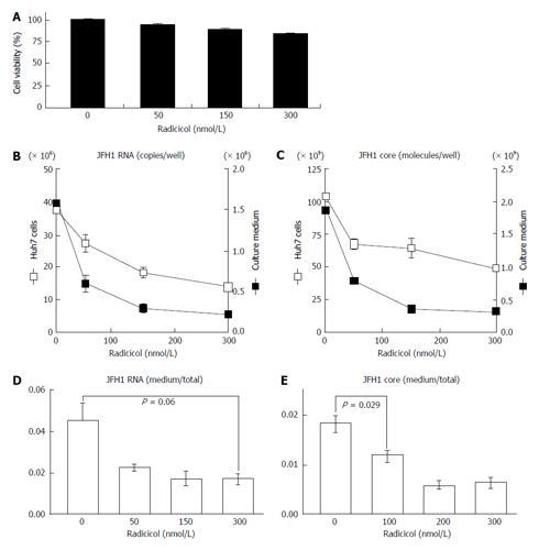
Figure 1 Radicicol affects the relative level of hepatitis C virus (core and hepatitis C virus RNA) produced from the JFH1/cell culture-derived hepatitis C virus system of Huh-7 cells.
A: After the cells were treated with radicicol (at final concentration of 0, 50, 150 and 300 nmol/L) for 12 h, the culture medium was replaced with fresh medium containing the same radicicol levels, and the HCV RNA and core levels released into the medium for the next 24 h and produced within cells were determined as described in the text. The cytotoxic effects of the treatment of radicicol on HCVcc Huh-7 cells were examined as described in the text; B and C: The levels of HCV RNA (B) and core (C) in the HCVcc Huh-7 cells (open squares) and the culture medium (filled squares) were quantified. The scales on the left sides (B and C) indicate the scales for the HCV RNA and the core in the Huh-7 cells. The scales on the right sides (B and C) indicate the scales for the HCV RNA and the core in the culture medium; D: The ratios of HCV RNA in the medium to the total HCV RNA (the sum of HCV RNA in the medium and in the cells) are shown; E: The ratios of the core in the medium to the total core (the sum of the core in the medium and in the cells) are shown. The data represent the mean values (± SEM) of the results from three independent experiments. HCV: Hepatitis C virus; HCVcc: Cell culture-derived HCV.
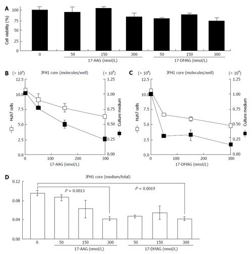
Figure 2 Effects of the geldanamycin derivatives 17-allylamino-17-demethoxygeldanamycin and 17-dimethylaminoethylamino-17-demethoxygeldanamycin on the release of JFH1.
A: The cytotoxic effects of 17-AAG and 17-DMAG on Huh-7 cells carrying JFH1/HCVcc were examined as described in the text; B and C: JFH1-infected Huh-7 cells were treated with 17-AAG (B) or 17-DMAG (C) for 24 h and then the protein samples were isolated and quantified. The levels of the HCV core present in the JFH1-infected Huh-7 cells (open squares) and the culture medium (filled squares) were examined as described in the text. The scales on the left (B and C) and right sides (B and C) indicate the scales for the HCV core in the Huh-7 cells and in the culture medium, respectively; D: The ratios of the core in the medium to the total core are shown. The concentrations of 17-AAG and 17-DMAG (0-300 nmol/L) are shown under the histograms. The data represent the mean values (± SEM) of the results from three independent experiments. HCV: Hepatitis C virus; 17-AGG: 17-allylamino-17-demethoxygeldanamycin; 17-DMAG: 17-dimethylaminoethylamino-17-demethoxygeldanamycin; HCVcc: Cell culture-derived HCV.
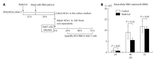
Figure 3 Infectivity of hepatitis C virus produced from Huh-7 cells that were treated with radicicol.
A: Experimental design. To examine whether the infectivity of the JFH1/HCVcc released from the Huh-7 cells in the presence of radicicol was altered, we infected fresh Huh-7 cells with JFH1/HCVcc prepared in the presence of radicicol. To prepare a viral stock, the JFH1/HCVcc infected Huh-7 cells with 50 nmol/L radicicol were maintained for an additional 36 h. HCVcc released in the medium during the last 24 h was collected and used as the viral stock. We diluted the viral stock 59.7 times (None) and 28.6 times (Radicicol) to reduce the effects of radicicol carryover and to adjust the HCV levels (core protein level: 200 fmols). The final radicicol concentration was 1.75 nmol/L which did not affect HCV propagation. The HCV samples were added to fresh Huh-7 cells and cultured for 24, 48 and 72 h. The HCV RNA in the infected Huh-7 cells was quantified as described in the text. The multiplicities of infection for HCV were 0.2 copies/cell and 0.16 copies/cell for the HCV infection without and with radicicol, respectively; B: The copies of HCV RNA in the Huh-7 cells of each well after infection (24, 48 and 48 h) were determined as described in the text. The value of the RNA copy number in the Huh-7 cells [which were infected with the viral stock from radicicol-treated cells (values of the closed bars)] was adjusted by multiplying by a factor of 1.25. The data represent the mean values (± SE) of the results from three independent experiments. HCV: Hepatitis C virus; HCVcc: Cell culture-derived HCV.
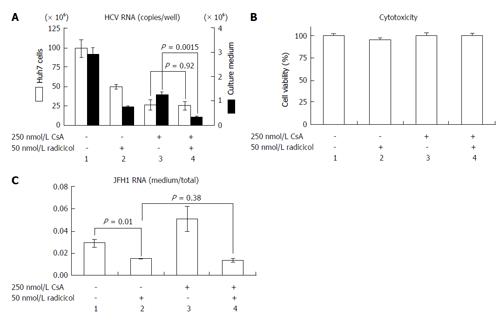
Figure 4 Radicicol and cyclosporin A have a synergistic effect on hepatitis C virus release into the medium.
A: JFH1/HCVcc-infected Huh-7 cells were treated with 250 nmol/L CsA alone, 50 nmol/L radicicol alone, or both for 36 h. The total RNA was prepared from the Huh-7 cells, and the HCV RNA was quantified by reverse transcription-polymerase chain reaction (open columns, the scale on the left of the panel indicates the copies/well). The medium was replaced by the fresh medium containing the same levels of the drugs after 12 h, and total RNA was prepared from the medium 24 h later. The copy numbers of the HCV RNA in the medium are indicated by the filled columns (scale on the right of the panel); B: The cytotoxic effects of the drugs used in (A) on the Huh-7 cells carrying JFH1/HCVcc; C: The ratios of the RNA in the medium to the total RNA (as described above) are shown. HCV: Hepatitis C virus; HCVcc: Cell culture-derived HCV; CsA: Cyclosporin A.
Figure 5 Radicicol and interferon-α have a synergistic effect on hepatitis C virus release into the medium.
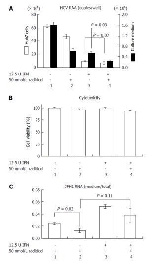
A: JFH1/HCVcc-infected Huh-7 cells were treated with 12.5 U INF-α alone, 50 nmol/L radicicol alone, or both for 36 h. The total RNA was prepared from the Huh-7 cells, and the HCV RNA was quantified by reverse transcription-polymerase chain reaction (open columns, the scale on the left of the panel indicates the copies/well). The medium was replaced by fresh medium after 12 h, and the total RNA was prepared from the medium 24 h later. The copy numbers of the HCV RNA in the medium are indicated by the filled columns (scale on the right of the panel); B: The cytotoxic effects of the drugs used in (A) on the Huh-7 cells carrying JFH1/HCVcc; C: The ratios of RNA in the medium to RNA in the total RNA (as described above) are shown. HCV: Hepatitis C virus; HCVcc: Cell culture-derived HCV; INF-α: Interferon-α.
Figure 6 Analysis of the synergistic effect of cyclosporin A and radicicol.
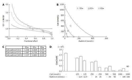
A: The graphs were constructed using the Chou and Talalay method. The CI and the fractional effect were derived from the release of the HCV core protein into the culture medium of JFH1-infected Huh-7 cells that were treated with a combination of CsA and radicicol (5:1 molar ratio). The open circles indicate actual experimental data. The CI vs fractional effect plots were generated with CalcSyn software. The dotted line and solid lines represent the mean values and standard deviation (1.96), respectively, of three independent experiments. The CI < 1, CI = 1, and CI > 1 indicate synergy, an additive effect and antagonism, respectively; B: The conservation isobologram (CI = 1) depicting different effective doses that yielded 50% (ED50), 75% (ED75) and 90% (ED90) inhibition of viral release by the combination treatment was graphed with actual experimental data (ED50, open triangles; ED75, open squares; ED90, open circles). The data represent the mean values of the results from three independent experiments; C: The CI values of each ED50, ED75 and ED90 by the combined CsA and radicicol treatment at molar ratios of 3:1, 5:1 and 10:1, respectively; D: The combined effect of CsA and radicicol at a molar ratio of 10:1. The HCV core levels in the medium are shown. The mean values and standard deviation (± SD) of the amounts of the HCV core released into the medium during three independent experiments are shown. The concentrations used in each set of experiments are shown under the histogram. ND: Not detected; CsA: Cyclosporin A; HCV: Hepatitis C virus; CI: Combination index.
- Citation: Kubota N, Nomoto M, Hwang GW, Watanabe T, Kohara M, Wakita T, Naganuma A, Kuge S. Hepatitis C virus inhibitor synergism suggests multistep interactions between heat-shock protein 90 and hepatitis C virus replication. World J Hepatol 2016; 8(5): 282-290
- URL: https://www.wjgnet.com/1948-5182/full/v8/i5/282.htm
- DOI: https://dx.doi.org/10.4254/wjh.v8.i5.282


