Copyright
©The Author(s) 2015.
World J Hepatol. Jul 28, 2015; 7(15): 1894-1898
Published online Jul 28, 2015. doi: 10.4254/wjh.v7.i15.1894
Published online Jul 28, 2015. doi: 10.4254/wjh.v7.i15.1894
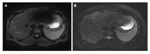
Figure 1 T2-weighted imaging with 3.
0 Tesla. In the absence of dedicated technical solutions, T2-weighted images at 3.0 Tesla (T) (A) are at risk of typical artefacts (namely standing-wave artefacts) causing signal drop-out over the field of view, especially left liver lobe. Focal liver lesions might be masked accordingly. The artefact is not present at 1.5 T (B).
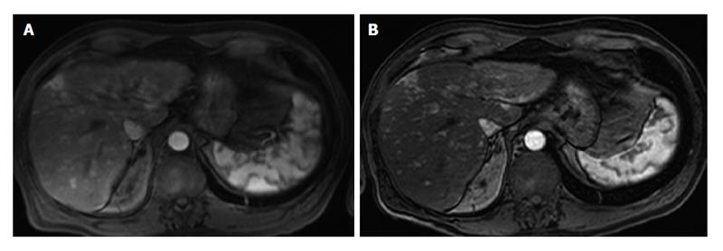
Figure 2 T1-weighted post-contrast imaging at 3.
0 Tesla. Compared to 1.5 Tesla (T) (A), post-contrast images acquired on 3.0 T magnets (B) show sharper details, as exemplified in this patient with chronic liver disease showing multiple artero-portal shunts.
- Citation: Girometti R. 3.0 Tesla magnetic resonance imaging: A new standard in liver imaging? World J Hepatol 2015; 7(15): 1894-1898
- URL: https://www.wjgnet.com/1948-5182/full/v7/i15/1894.htm
- DOI: https://dx.doi.org/10.4254/wjh.v7.i15.1894


