Copyright
©2014 Baishideng Publishing Group Co.
World J Hepatol. Apr 27, 2014; 6(4): 251-262
Published online Apr 27, 2014. doi: 10.4254/wjh.v6.i4.251
Published online Apr 27, 2014. doi: 10.4254/wjh.v6.i4.251
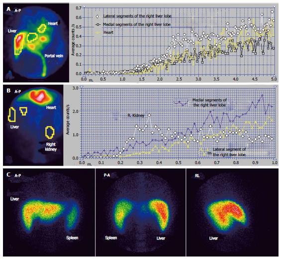
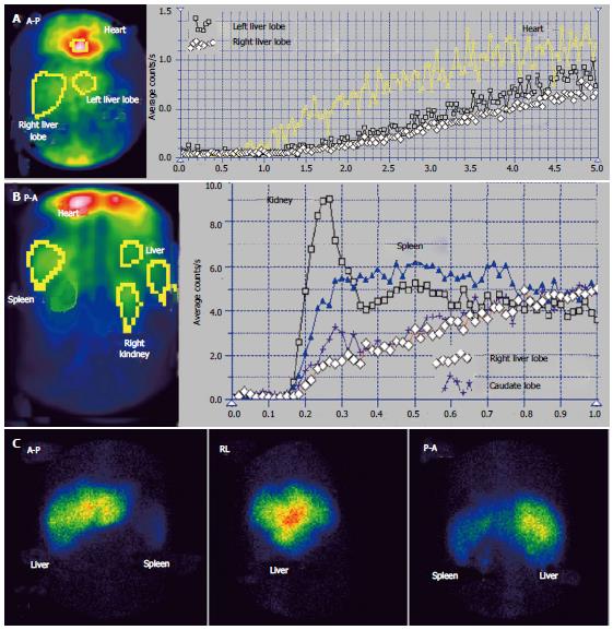
Figure 1 Budd-Chiari syndrome with obstruction of the middle hepatic vein.
A: Per-rectal portal scintigraphy; B: Liver angioscintigraphy; C: Liver scan.
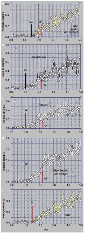
Figure 2 Per-rectal portal scintigraphy in Budd-Chiari syndrome with obstruction of the middle hepatic vein.
A: Curve on the territory drained by the middle hepatic vein; B: Caudate lobe curve; C: Left lobe curve; D: Curve on the territory drained by the right hepatic vein; E: Heart curve.
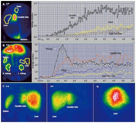
Figure 3 Budd-Chiari syndrome with old obstruction of the left hepatic vein and recent obstruction of the middle and right hepatic veins.
A: Per-rectal portal scintigraphy; B: Liver angioscintigraphy; C: Liver scan.
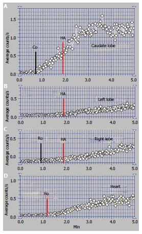
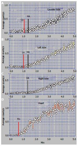
Figure 4 Per-rectal portal scintigraphy in Budd-Chiari syndrome with old obstruction of the left hepatic vein and recent obstruction of the middle and right hepatic veins.
A: Caudate lobe curve; B: Left lobe curve; C: Right lobe curve; D: Heart curve.
Figure 5 Budd-Chiari syndrome with old obstruction of the middle and right hepatic veins also followed by old obstruction of the left hepatic vein.
A: Per-rectal portal scintigraphy; B: Liver angioscintigraphy; C: Liver scan.
Figure 6 Per-rectal portal scintigraphy in Budd-Chiari syndrome with old obstruction of the middle and right hepatic veins also followed by old obstruction of the left hepatic vein.
A: Caudate lobe curve; B: Left lobe curve; C: Right lobe curve; D: Heart curve.
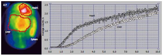
Figure 7 Per-rectal portal scintigraphy of a patient with Budd-Chiari syndrome with old obstruction of the middle and right hepatic veins and recent obstruction of the left hepatic vein.
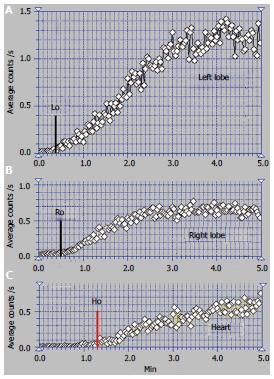
Figure 8 Per-rectal portal scintigraphy in Budd-Chiari syndrome with obstruction of the terminal part of the inferior vena cava.
A: Left lobe curve; B: Right lobe curve; C: Heart curve.
- Citation: Dragoteanu M, Balea IA, Piglesan CD. Nuclear medicine dynamic investigations in the diagnosis of Budd-Chiari syndrome. World J Hepatol 2014; 6(4): 251-262
- URL: https://www.wjgnet.com/1948-5182/full/v6/i4/251.htm
- DOI: https://dx.doi.org/10.4254/wjh.v6.i4.251


