Copyright
©The Author(s) 2020.
World J Hepatol. Aug 27, 2020; 12(8): 519-524
Published online Aug 27, 2020. doi: 10.4254/wjh.v12.i8.519
Published online Aug 27, 2020. doi: 10.4254/wjh.v12.i8.519
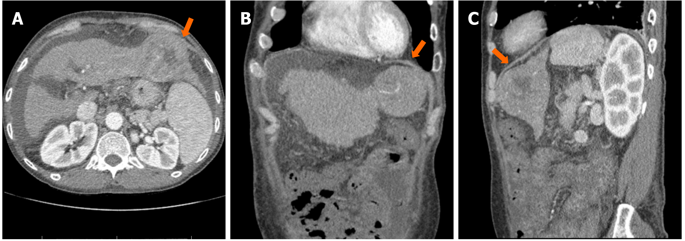
Figure 1 Arterial phase of abdominal computed tomography showed a centrally necrotic mass in the left hepatic lobe, measuring 6.
7 cm × 6.5 cm (orange arrow). A: Axial view; B: Coronal view; C: Sagittal view.
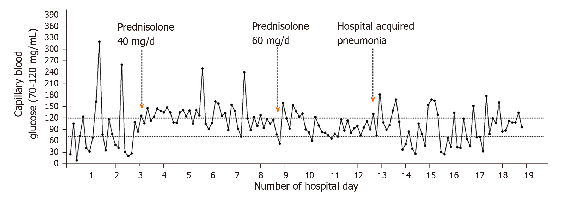
Figure 2 Capillary blood glucose trend.
- Citation: Yu B, Douli R, Suarez JA, Gutierrez VP, Aldiabat M, Khan M. Non-islet cell tumor hypoglycemia as an initial presentation of hepatocellular carcinoma coupled with end-stage liver cirrhosis: A case report and review of literature. World J Hepatol 2020; 12(8): 519-524
- URL: https://www.wjgnet.com/1948-5182/full/v12/i8/519.htm
- DOI: https://dx.doi.org/10.4254/wjh.v12.i8.519


