Copyright
©The Author(s) 2018.
World J Hepatol. Apr 27, 2018; 10(4): 402-408
Published online Apr 27, 2018. doi: 10.4254/wjh.v10.i4.402
Published online Apr 27, 2018. doi: 10.4254/wjh.v10.i4.402
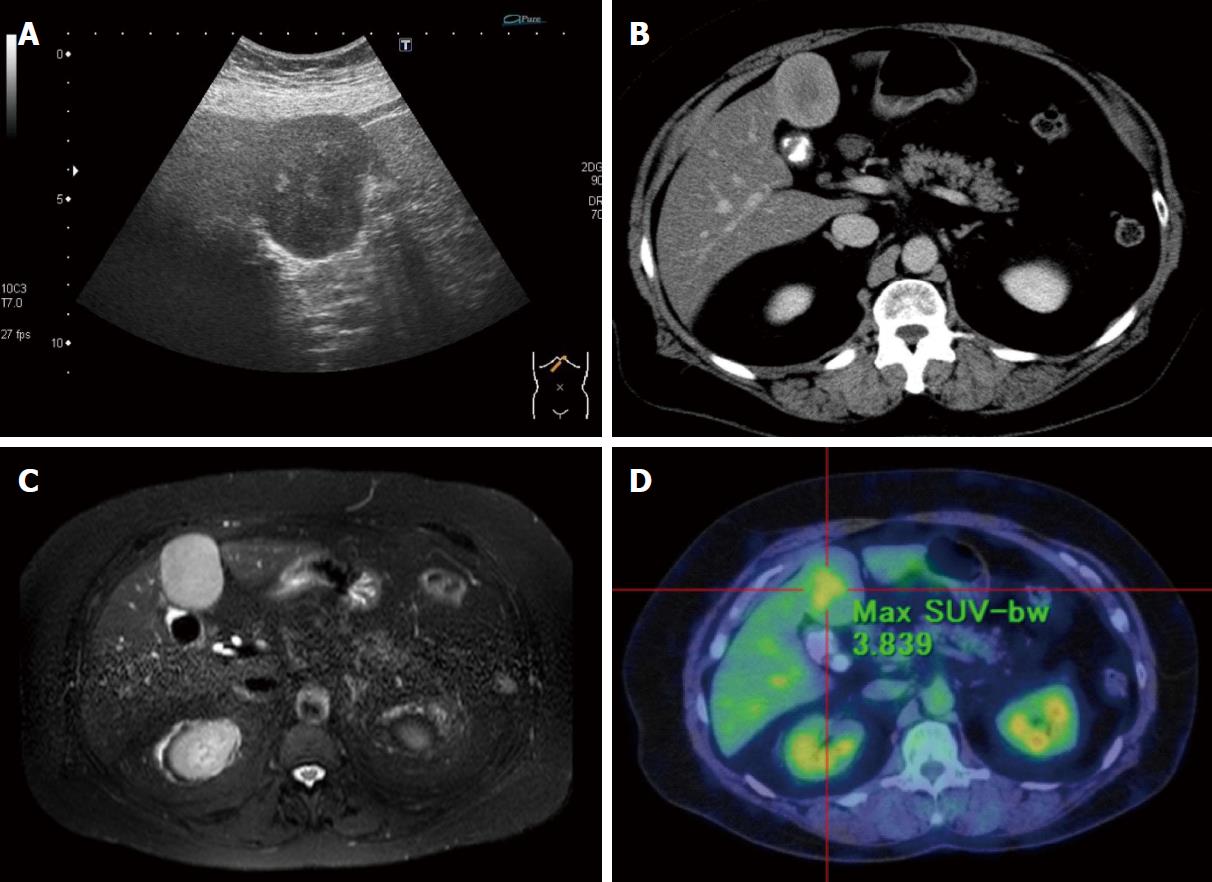
Figure 1 Evaluation of clinical findings.
A: Ultrasonography of the liver showed a very-low-echoic smooth mass (60 mm × 40 mm) with a heterogeneous high-echoic region in the median section of the left lobe of the liver (Segment 4); B: Axial delayed phase CE-CT showed the lesion to be gradually enhanced heterogeneously; C: The lesion showed a hyperintense heterogeneous region on axial T2-weighted EOB-MRI; D: The lesion showed obvious metabolically active foci by 18-fluorodeoxyglucose-PET-CT evaluation, while other lesions were not detected.
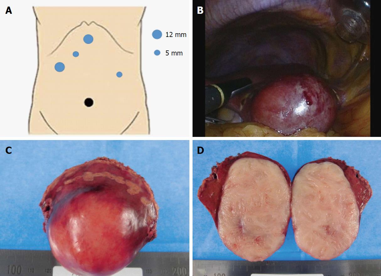
Figure 2 Ultrasound-guided pure laparoscopic partial liver resection (Segment 4) and surgical specimen.
A: Five ports were placed for liver partial resection. Three ports were placed around the epigastric region as working ports (blue circles). An umbilical port was used as the camera port (black circle). The resected partial liver was retrieved from the umbilical port with auxiliary incision. A Pringle maneuver was used for the left lateral port; B: We performed ultrasound-guided pure laparoscopic partial liver resection with vessel-sealing devices (LigaSure™ Maryland Jaw 37 cm Laparoscopic Sealer/Divider, Medtronic, Dublin, Ireland) using the crush clamping method and Pringle maneuver; C: Macroscopic image of the resected specimen showed a smooth surface mass; D: Cross-section of the resected specimen showed a milky-white-colored solid mass without necrotic lesion.
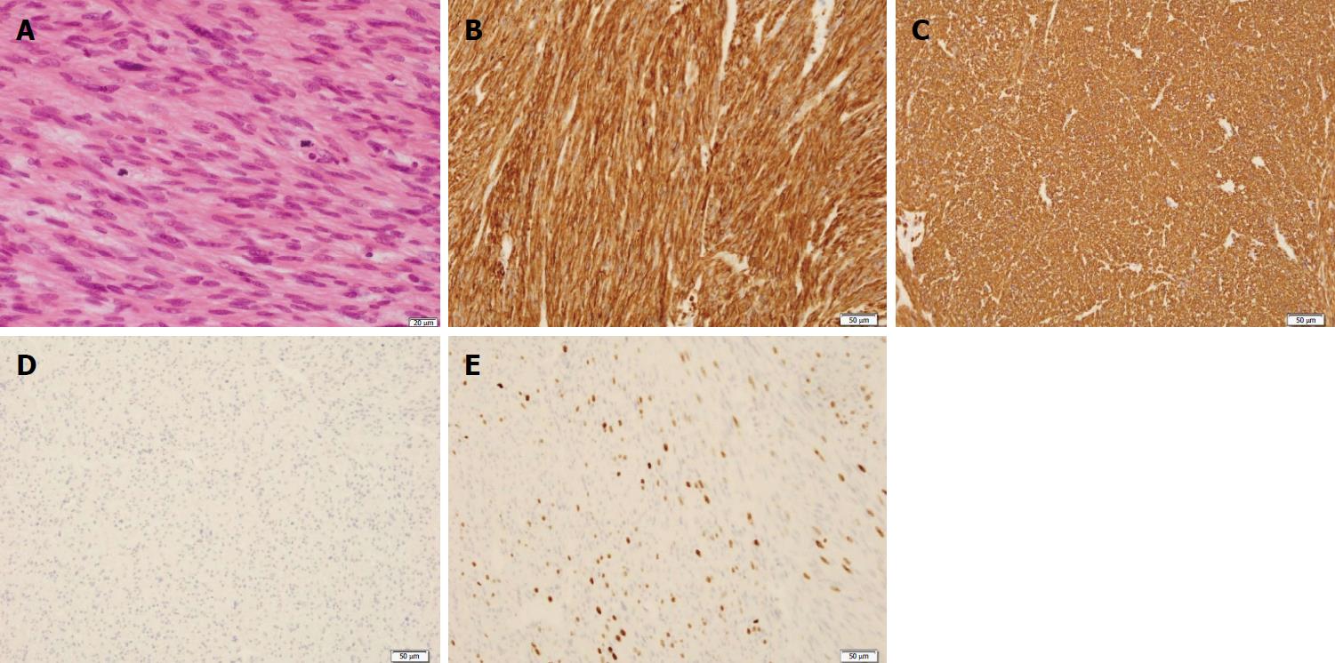
Figure 3 Pathological findings of the resected specimen.
A: HE stain showed the proliferation of spindle-shaped cells with ≥ 10 MFs-/-10 HPFs and diffuse moderate-to-severe atypia without coagulative tumor cell necrosis; B: Immunohistochemical findings were strongly positive for SMA; C: Immunohistochemical findings were strongly positive for desmin; D: Immunohistochemical findings were strongly positive for negative for c-kit; E: Approximately 35% ki-67 positive cells were seen in the lesion. Scale bars: 20 μm (A), 50 μm (B-E).
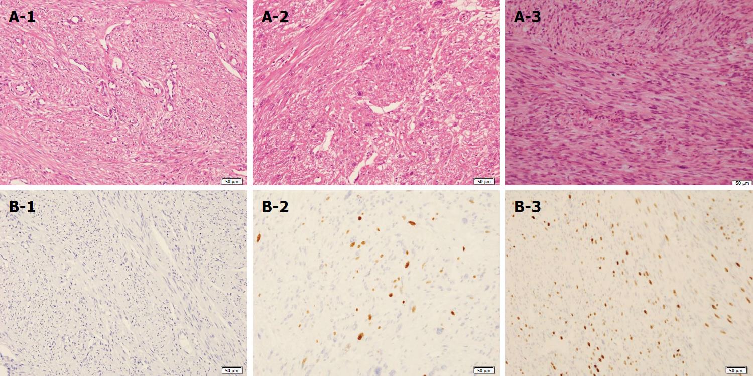
Figure 4 Pathological reconfirmation of uterine leiomyoma and broad ligament with STUMP and liver lesion with leiomyosarcoma.
MF was absent in an HE stain of the uterine leiomyoma (A-1). The number of MFs was 1-5 MFs / 10 HPFs in the broad ligament (A-2) and more than 10 MFs/10 HPFs in the liver mass (A-3). Ki-67-positive cells were not seen in the uterine leiomyoma (B-1). The MIB-1 index was approximately 5% of the broad ligament and approximately 35% of the liver mass (B-2) (B-3). The pathological findings of MFs and the MIB-1 index showed malignant transformation from STUMP to leiomyosarcoma. Scale bars: 50 μm.
- Citation: Fukui K, Takase N, Miyake T, Hisano K, Maeda E, Nishimura T, Abe K, Kozuki A, Tanaka T, Harada N, Takamatsu M, Kaneda K. Review of the literature laparoscopic surgery for metastatic hepatic leiomyosarcoma associated with smooth muscle tumor of uncertain malignant potential: Case report. World J Hepatol 2018; 10(4): 402-408
- URL: https://www.wjgnet.com/1948-5182/full/v10/i4/402.htm
- DOI: https://dx.doi.org/10.4254/wjh.v10.i4.402


