Copyright
©The Author(s) 2015.
World J Stem Cells. May 26, 2015; 7(4): 776-788
Published online May 26, 2015. doi: 10.4252/wjsc.v7.i4.776
Published online May 26, 2015. doi: 10.4252/wjsc.v7.i4.776
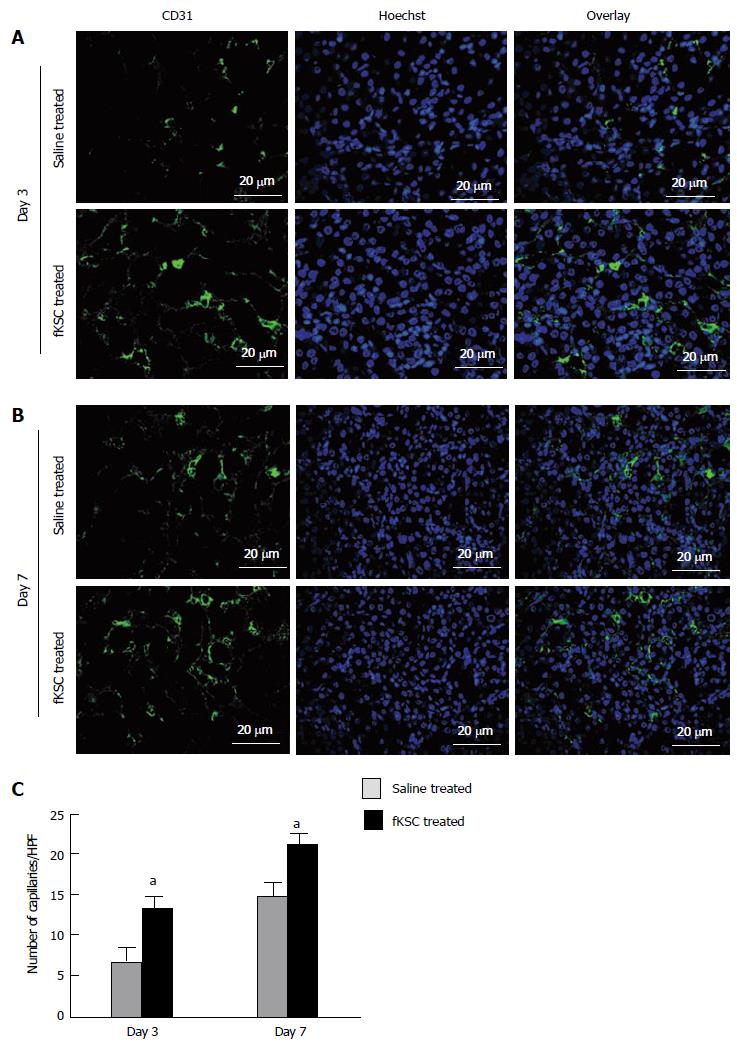
Figure 7 Fetal kidney stem cells accelerate angiogenesis in rats with cisplatin induced acute renal failure.
Representative immunoflourescent photomicrographs (Scale bars indicate 20 μm) showing perivascular capillaries stained with CD31 in saline treated and fetal kidney stem cells (fKSC) treated groups on day 3 (A) and on day 7 (B) after fKSC therapy. C: Quantification of perivascular capillaries stained with CD31, expressed as capillaries per HPF, in saline and fKSC treated groups. Values expressed Mean ± SE. aP < 0.05 for fKSC vs saline treated group. HPF: High-power field.
- Citation: Gupta AK, Jadhav SH, Tripathy NK, Nityanand S. Fetal kidney stem cells ameliorate cisplatin induced acute renal failure and promote renal angiogenesis. World J Stem Cells 2015; 7(4): 776-788
- URL: https://www.wjgnet.com/1948-0210/full/v7/i4/776.htm
- DOI: https://dx.doi.org/10.4252/wjsc.v7.i4.776


