Copyright
©The Author(s) 2015.
World J Stem Cells. May 26, 2015; 7(4): 776-788
Published online May 26, 2015. doi: 10.4252/wjsc.v7.i4.776
Published online May 26, 2015. doi: 10.4252/wjsc.v7.i4.776
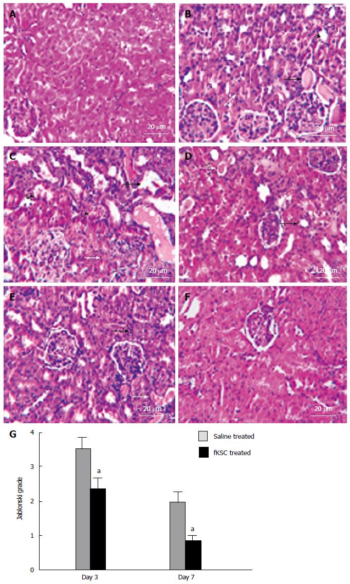
Figure 4 Fetal kidney stem cells attenuate renal structural damage in rats with cisplatin induced acute renal failure (Scale bars indicate 20 μm).
A: Kidney section of healthy control rats showing the normal architecture of tubules and glomeruli; B: Kidney section after 5 d of cisplatin treatment showing severe tubular necrosis (white arrow), hyaline cast formation (black arrow) and tubular dilatation (black arrow head); C: Kidney section of saline treated rats on day 3 of therapy showing severe tubular necrosis (white arrow), tubular vacuolization (black arrow) and tubular swelling and obstruction (black arrow head) with cell debris on day 3 after therapy; D: Kidney section of fetal kidney stem cells (fKSC) treated rats on day 3 of therapy showing attenuation of tubular injury with mild tubular dilatation (black arrow) and few hyaline casts inside the lumen (white arrow); E: Kidney section of saline treated rats on day 7 of therapy showing few necrotic tubular cells (black arrow) and luminal hyaline casts (white arrow); F: Kidney section of fKSC treated rats on day 7 of therapy fKSC showing almost normal architecture of tubules; G: Jablonski grading score of histological assessment of tubular necrosis in saline and fKSC treated kidneys on day 3 and day 7. Values expressed as mean ± SE. aP < 0.05 for fKSC vs saline treated group.
- Citation: Gupta AK, Jadhav SH, Tripathy NK, Nityanand S. Fetal kidney stem cells ameliorate cisplatin induced acute renal failure and promote renal angiogenesis. World J Stem Cells 2015; 7(4): 776-788
- URL: https://www.wjgnet.com/1948-0210/full/v7/i4/776.htm
- DOI: https://dx.doi.org/10.4252/wjsc.v7.i4.776


