Copyright
©2013 Baishideng Publishing Group Co.
World J Stem Cells. Jul 26, 2013; 5(3): 86-97
Published online Jul 26, 2013. doi: 10.4252/wjsc.v5.i3.86
Published online Jul 26, 2013. doi: 10.4252/wjsc.v5.i3.86
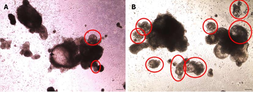
Figure 7 Phase contrast microscopy of cardiomyocytes obtained from differentiation of HES-3 cells.
Pictures of aggregates formed from human embryonic stem cells differentiated in SB media (A) and SupSB media (B) were taken after 16 d. Beating areas are indicated by red circles. Total number of beating aggregates was higher in cultures differentiated in SupSB medium(about 80% of total aggregates) compared to in SB medium (about 60% of total aggregates). Scale bar = 200 μm.
- Citation: Ting S, Lecina M, Chan YC, Tse HF, Reuveny S, Oh SK. Nutrient supplemented serum-free medium increases cardiomyogenesis efficiency of human pluripotent stem cells. World J Stem Cells 2013; 5(3): 86-97
- URL: https://www.wjgnet.com/1948-0210/full/v5/i3/86.htm
- DOI: https://dx.doi.org/10.4252/wjsc.v5.i3.86


