Copyright
©2013 Baishideng Publishing Group Co.
World J Stem Cells. Apr 26, 2013; 5(2): 43-52
Published online Apr 26, 2013. doi: 10.4252/wjsc.v5.i2.43
Published online Apr 26, 2013. doi: 10.4252/wjsc.v5.i2.43
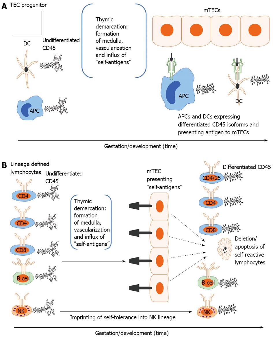
Figure 2 Developmental acquisition of self-tolerance: Antigen exposure model.
A: Thymic epithelial (TEC) progenitors mature in intimate contact with dendritic cells (DCs) and likely other antigen presenting cells (APCs) thus forming the thymic medulla (i.e., thymic demarcation). With vascularization, self-antigens (i.e., antigens present during the finite tolerance window) from the periphery are processed and presented to medullary thymic epithelial cells (mTECs). The developing mTEC matrix is only receptive to antigen presentation during the tolerance window (time dependence) and subsequently exhibits on its surface the self-antigen repertoire (see 3. Ramifications: Dissection of immune tolerance); B: All lymphoid compartments [T, B and natural killer (NK) cell] enter the thymus expressing undifferentiated CD45 but are lineage defined. They then migrate through the thymus encountering mTECs expressing the self-antigen repertoire. CD4, CD8 and B cells reactive to self-antigens are deleted. CD4/CD25 cells reactive to self-antigens are rendered suppressive (Treg) in Hassal’s corpuscles. NK cells are imprinted in an undefined fashion (likely based on major histocompatibility complex exposure) to become tolerant to self. All differentiated lymphocytes (i.e., cells that have undergone CD45 isoform maturation and are not self-reactive or are tolerogenic (i.e.,Treg) then migrate to the spleen or other secondary lymphoid organs (see 3. Ramifications: Dissection of self-tolerance)[6,21,31-37,42].
- Citation: Pixley JS, Zanjani ED. In utero transplantation: Disparate ramifications. World J Stem Cells 2013; 5(2): 43-52
- URL: https://www.wjgnet.com/1948-0210/full/v5/i2/43.htm
- DOI: https://dx.doi.org/10.4252/wjsc.v5.i2.43


