Copyright
©The Author(s) 2015.
World J Stem Cells. Jan 26, 2015; 7(1): 75-83
Published online Jan 26, 2015. doi: 10.4252/wjsc.v7.i1.75
Published online Jan 26, 2015. doi: 10.4252/wjsc.v7.i1.75
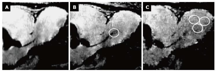
Figure 1 Monitoring the migration of endogenous neural stem cell labeled with iron oxide particles in the rat brain in vivo, using magnetic resonance imaging.
Migrating cells were imaged 1 d (A), 3 d (B), and 8 d (C) after labeling, migrating from the subventricular zone (left in the sagittal images) to the olfactory bulb (on the right). White circles surround migrating cells. Granot et al[54], with permission. MRI: Magnetic resonance imaging.
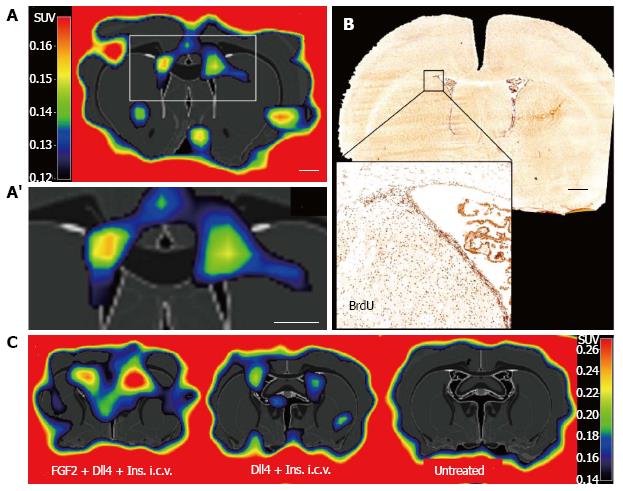
Figure 2 [18F]FLT labels proliferating endogenous neural stem cell in the neurogenic niches of the healthy rodent brain.
A, A’: eNSC proliferation in the subventricular zone of adult rats as assessed by [18F]FLT-PET; B: The signal corresponds to BrdU-positive cells in the region; C: Activation of eNSC by pharmacological stimulation with fibroblast growth factor 2, delta-like 4, and insulin is visualized with [18F]FLT-PET. eNSC: Endogenous neural stem cell; [18F]FLT-PET: 3’-deoxy-3’-[18F]fluoro-L-thymidine-positron emission tomography; i.c.v.: Intracerebroventricular application. Adapted from Rueger et al[75], with permission.
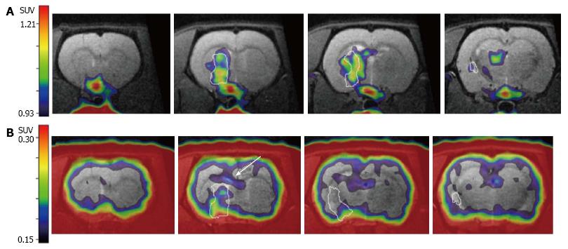
Figure 3 Conclusive differentiation between cell proliferation from endogenous neural stem cell and immune cells.
After transient focal ischemia (lesion outlined in white), A: Neuroinflammatory processes are visualized with [11C]PK11195-PET, depicting inflammation in the infarct core as well as in the ischemic border zone; B: [18F]FLT-PET data acquired in the same imaging session without moving the animals in the scanner demonstrates additional cell proliferation in the subventricular zone (white arrow), originating from endogenous neural stem cells. Adapted from Rueger et al[97], with permission. [18F]FLT-PET: 3’-deoxy-3’-[18F]fluoro-L-thymidine-positron emission tomography.
-
Citation: Rueger MA, Schroeter M.
In vivo imaging of endogenous neural stem cells in the adult brain. World J Stem Cells 2015; 7(1): 75-83 - URL: https://www.wjgnet.com/1948-0210/full/v7/i1/75.htm
- DOI: https://dx.doi.org/10.4252/wjsc.v7.i1.75


