Copyright
©The Author(s) 2015.
World J Stem Cells. Jan 26, 2015; 7(1): 75-83
Published online Jan 26, 2015. doi: 10.4252/wjsc.v7.i1.75
Published online Jan 26, 2015. doi: 10.4252/wjsc.v7.i1.75
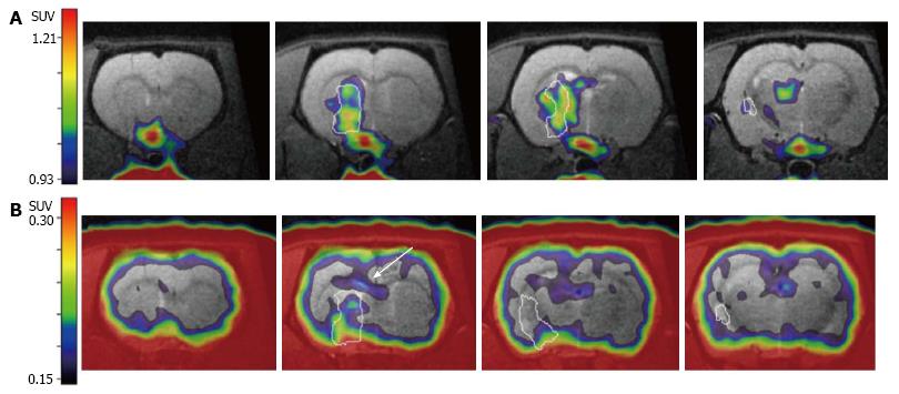
Figure 3 Conclusive differentiation between cell proliferation from endogenous neural stem cell and immune cells.
After transient focal ischemia (lesion outlined in white), A: Neuroinflammatory processes are visualized with [11C]PK11195-PET, depicting inflammation in the infarct core as well as in the ischemic border zone; B: [18F]FLT-PET data acquired in the same imaging session without moving the animals in the scanner demonstrates additional cell proliferation in the subventricular zone (white arrow), originating from endogenous neural stem cells. Adapted from Rueger et al[97], with permission. [18F]FLT-PET: 3’-deoxy-3’-[18F]fluoro-L-thymidine-positron emission tomography.
-
Citation: Rueger MA, Schroeter M.
In vivo imaging of endogenous neural stem cells in the adult brain. World J Stem Cells 2015; 7(1): 75-83 - URL: https://www.wjgnet.com/1948-0210/full/v7/i1/75.htm
- DOI: https://dx.doi.org/10.4252/wjsc.v7.i1.75


