Copyright
©2012 Baishideng.
World J Stem Cells. Sep 26, 2012; 4(9): 94-100
Published online Sep 26, 2012. doi: 10.4252/wjsc.v4.i9.94
Published online Sep 26, 2012. doi: 10.4252/wjsc.v4.i9.94
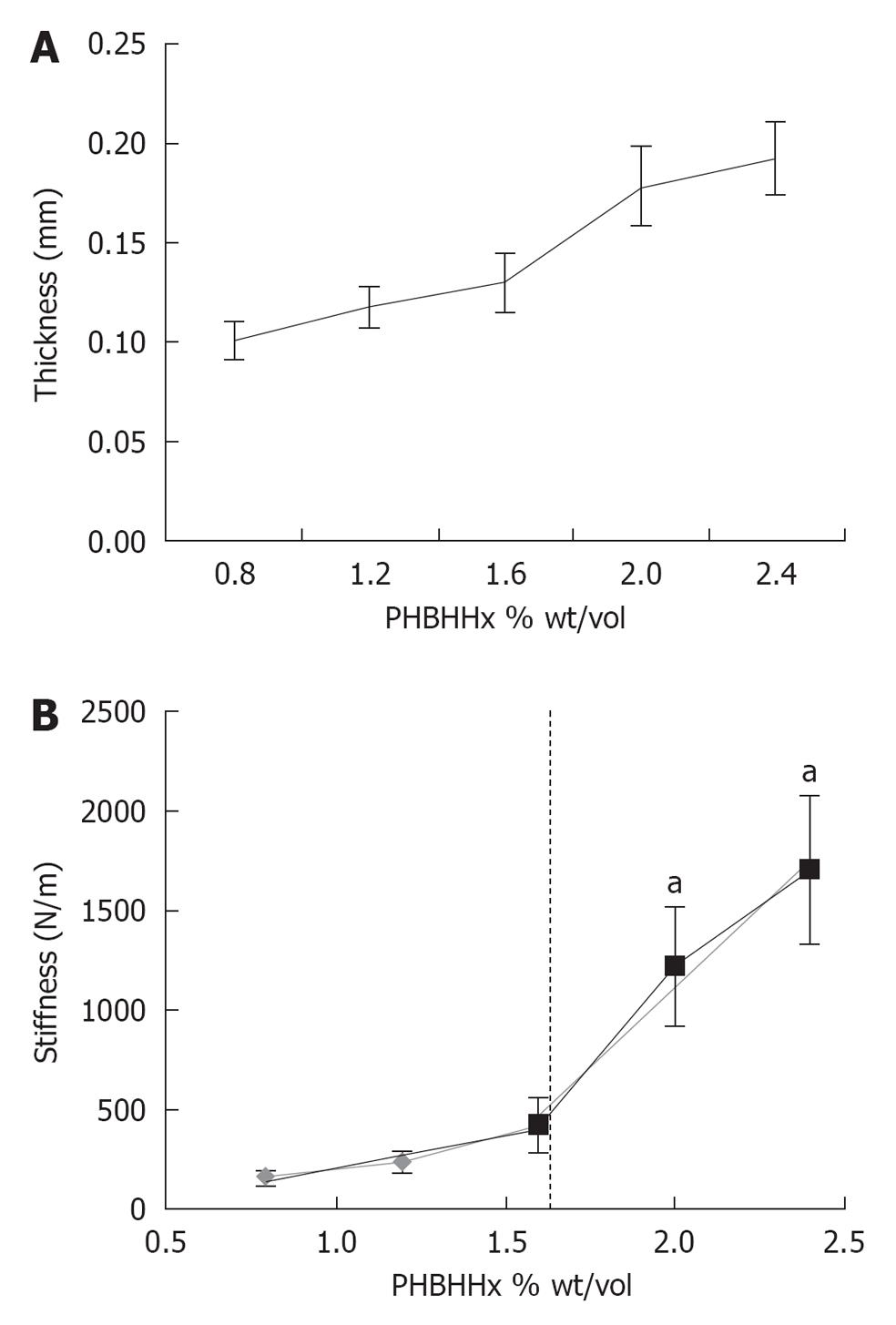
Figure 1 Characterization of poly(3-hydroxybutyrate-co-3-hydroxyhexanoate) films.
A: Optical Coherence Tomography and Image J software analysis were used to determine poly(3-hydroxybutyrate-co-3-hydroxyhexanoate) (PHBHHx) film thickness. Average ± 1SD shown on graph, n = 9; B: PHBHHX film stiffness was measured with the BOSE ElectoForce 3200 system. Average ± 1 SD shown on graph, n = 3. aIndicates significant increase compared to ≤ 0.6% weight/volume PHBHHx. Trend lines are indicated by hatched red lines.
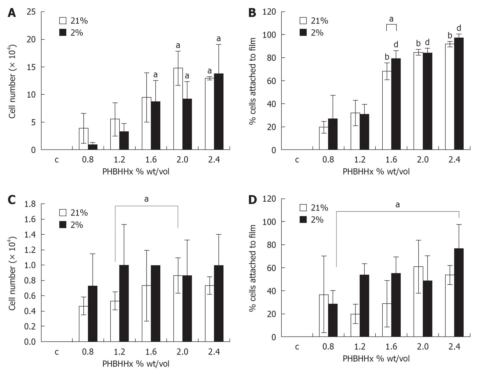
Figure 2 Cell attachment to poly(3-hydroxybutyrate-co-3-hydroxyhexanoate) films.
A: Number of tenocytes attached to poly(3-hydroxybutyrate-co-3-hydroxyhexanoate) (PHBHHx) films of varying % weight/volume polymer concentration; B: Tenocyte attachment to films of varying % weight/volume polymer concentration as a percentage of total cell number in the well; C: Number of human mesenchymal stem cells (hMSCs) attached to varying % weight/volume concentration of polymer; D: hMSCs attachment to films of varying % weight/volume polymer concentration as a percentage of total cell number in the well. Axes are as labeled, error bars indicate one standard deviation. aIndicates P≤ 0.05, bP≤ 0.02, dP≤ 0.01 vs≤ 0.8% weight/volume PHBHHx or as indicated.
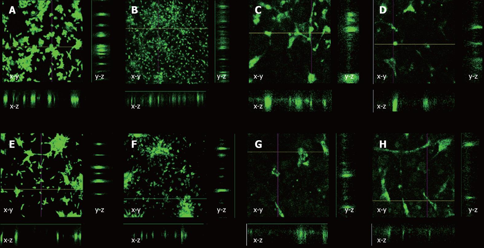
Figure 3 Representative images showing surface and cross section views through poly(3-hydroxybutyrate-co-3-hydroxyhexanoate) films.
A: tenocytes 21% O2, 24 h; B: tenocytes 21% O2, 72 h; C: Human mesenchymal stem cells (hMSCs) 21% O2, 24 h; D: hMSCs 21% O2, 72 h; E: Tenocytes 2% O2, 24 h; F: Tenocytes 2% O2, 72 h; G: hMSCs 2% O2, 24 h; H: hMSCs 2% O2, 72 h. All images were taken at 10 × magnification. x-y indicates surface view, x-z and y-z indicate reconstructed cross section views.
- Citation: Lomas AJ, Chen GG, El Haj AJ, Forsyth NR. Poly(3-hydroxybutyrate-co-3-hydroxyhexanoate) supports adhesion and migration of mesenchymal stem cells and tenocytes. World J Stem Cells 2012; 4(9): 94-100
- URL: https://www.wjgnet.com/1948-0210/full/v4/i9/94.htm
- DOI: https://dx.doi.org/10.4252/wjsc.v4.i9.94


